Sodium »
PDB 2r25-2uyz »
2ra9 »
Sodium in PDB 2ra9: Crystal Structure of A DUF1285 Family Protein (SBAL_2486) From Shewanella Baltica OS155 at 1.40 A Resolution
Protein crystallography data
The structure of Crystal Structure of A DUF1285 Family Protein (SBAL_2486) From Shewanella Baltica OS155 at 1.40 A Resolution, PDB code: 2ra9
was solved by
Joint Center For Structural Genomics (Jcsg),
with X-Ray Crystallography technique. A brief refinement statistics is given in the table below:
| Resolution Low / High (Å) | 29.85 / 1.40 |
| Space group | P 21 21 21 |
| Cell size a, b, c (Å), α, β, γ (°) | 38.409, 62.286, 73.247, 90.00, 90.00, 90.00 |
| R / Rfree (%) | 16.2 / 19.6 |
Sodium Binding Sites:
The binding sites of Sodium atom in the Crystal Structure of A DUF1285 Family Protein (SBAL_2486) From Shewanella Baltica OS155 at 1.40 A Resolution
(pdb code 2ra9). This binding sites where shown within
5.0 Angstroms radius around Sodium atom.
In total only one binding site of Sodium was determined in the Crystal Structure of A DUF1285 Family Protein (SBAL_2486) From Shewanella Baltica OS155 at 1.40 A Resolution, PDB code: 2ra9:
In total only one binding site of Sodium was determined in the Crystal Structure of A DUF1285 Family Protein (SBAL_2486) From Shewanella Baltica OS155 at 1.40 A Resolution, PDB code: 2ra9:
Sodium binding site 1 out of 1 in 2ra9
Go back to
Sodium binding site 1 out
of 1 in the Crystal Structure of A DUF1285 Family Protein (SBAL_2486) From Shewanella Baltica OS155 at 1.40 A Resolution
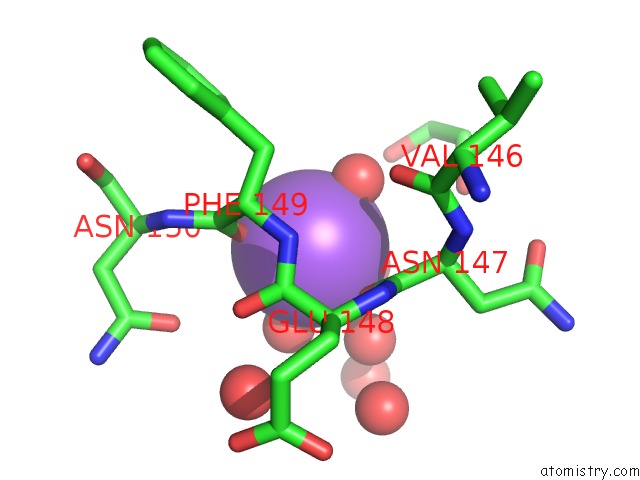
Mono view
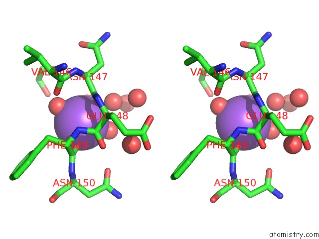
Stereo pair view
Mono view
Stereo pair view
A full contact list of Sodium with other atoms in the Na binding
site number 1 of Crystal Structure of A DUF1285 Family Protein (SBAL_2486) From Shewanella Baltica OS155 at 1.40 A Resolution within 5.0Å range:
|
Reference:
G.W.Han,
C.Bakolitsa,
M.D.Miller,
A.Kumar,
D.Carlton,
R.J.Najmanovich,
P.Abdubek,
T.Astakhova,
H.L.Axelrod,
C.Chen,
H.J.Chiu,
T.Clayton,
D.Das,
M.C.Deller,
L.Duan,
D.Ernst,
J.Feuerhelm,
J.C.Grant,
A.Grzechnik,
L.Jaroszewski,
K.K.Jin,
H.A.Johnson,
H.E.Klock,
M.W.Knuth,
P.Kozbial,
S.S.Krishna,
D.Marciano,
D.Mcmullan,
A.T.Morse,
E.Nigoghossian,
L.Okach,
R.Reyes,
C.L.Rife,
N.Sefcovic,
H.J.Tien,
C.B.Trame,
H.Van Den Bedem,
D.Weekes,
Q.Xu,
K.O.Hodgson,
J.Wooley,
M.A.Elsliger,
A.M.Deacon,
A.Godzik,
S.A.Lesley,
I.A.Wilson.
Structures of the First Representatives of Pfam Family PF06938 (DUF1285) Reveal A New Fold with Repeated Structural Motifs and Possible Involvement in Signal Transduction. Acta Crystallogr.,Sect.F V. 66 1218 2010.
ISSN: ESSN 1744-3091
PubMed: 20944214
DOI: 10.1107/S1744309109050416
Page generated: Mon Oct 7 04:20:31 2024
ISSN: ESSN 1744-3091
PubMed: 20944214
DOI: 10.1107/S1744309109050416
Last articles
Zn in 9MJ5Zn in 9HNW
Zn in 9G0L
Zn in 9FNE
Zn in 9DZN
Zn in 9E0I
Zn in 9D32
Zn in 9DAK
Zn in 8ZXC
Zn in 8ZUF