Sodium »
PDB 2qd8-2r22 »
2qz2 »
Sodium in PDB 2qz2: Crystal Structure of A Glycoside Hydrolase Family 11 Xylanase From Aspergillus Niger in Complex with Xylopentaose
Enzymatic activity of Crystal Structure of A Glycoside Hydrolase Family 11 Xylanase From Aspergillus Niger in Complex with Xylopentaose
All present enzymatic activity of Crystal Structure of A Glycoside Hydrolase Family 11 Xylanase From Aspergillus Niger in Complex with Xylopentaose:
3.2.1.8;
3.2.1.8;
Protein crystallography data
The structure of Crystal Structure of A Glycoside Hydrolase Family 11 Xylanase From Aspergillus Niger in Complex with Xylopentaose, PDB code: 2qz2
was solved by
E.Vandermarliere,
S.Rombouts,
S.V.Strelkov,
J.A.Delcour,
C.M.Courtin,
A.Rabijns,
with X-Ray Crystallography technique. A brief refinement statistics is given in the table below:
| Resolution Low / High (Å) | 19.48 / 2.80 |
| Space group | P 65 2 2 |
| Cell size a, b, c (Å), α, β, γ (°) | 67.326, 67.326, 165.401, 90.00, 90.00, 120.00 |
| R / Rfree (%) | 22.1 / 27.7 |
Sodium Binding Sites:
The binding sites of Sodium atom in the Crystal Structure of A Glycoside Hydrolase Family 11 Xylanase From Aspergillus Niger in Complex with Xylopentaose
(pdb code 2qz2). This binding sites where shown within
5.0 Angstroms radius around Sodium atom.
In total only one binding site of Sodium was determined in the Crystal Structure of A Glycoside Hydrolase Family 11 Xylanase From Aspergillus Niger in Complex with Xylopentaose, PDB code: 2qz2:
In total only one binding site of Sodium was determined in the Crystal Structure of A Glycoside Hydrolase Family 11 Xylanase From Aspergillus Niger in Complex with Xylopentaose, PDB code: 2qz2:
Sodium binding site 1 out of 1 in 2qz2
Go back to
Sodium binding site 1 out
of 1 in the Crystal Structure of A Glycoside Hydrolase Family 11 Xylanase From Aspergillus Niger in Complex with Xylopentaose
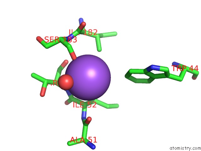
Mono view
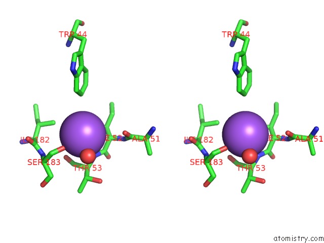
Stereo pair view
Mono view
Stereo pair view
A full contact list of Sodium with other atoms in the Na binding
site number 1 of Crystal Structure of A Glycoside Hydrolase Family 11 Xylanase From Aspergillus Niger in Complex with Xylopentaose within 5.0Å range:
|
Reference:
E.Vandermarliere,
T.M.Bourgois,
S.Rombouts,
S.Van Campenhout,
G.Volckaert,
S.V.Strelkov,
J.A.Delcour,
A.Rabijns,
C.M.Courtin.
Crystallographic Analysis Shows Substrate Binding at the -3 to +1 Active-Site Subsites and at the Surface of Glycoside Hydrolase Family 11 Endo-1,4-Beta-Xylanases. Biochem.J. V. 410 71 2008.
ISSN: ISSN 0264-6021
PubMed: 17983355
DOI: 10.1042/BJ20071128
Page generated: Mon Oct 7 04:06:18 2024
ISSN: ISSN 0264-6021
PubMed: 17983355
DOI: 10.1042/BJ20071128
Last articles
Zn in 9J0NZn in 9J0O
Zn in 9J0P
Zn in 9FJX
Zn in 9EKB
Zn in 9C0F
Zn in 9CAH
Zn in 9CH0
Zn in 9CH3
Zn in 9CH1