Sodium »
PDB 2qd8-2r22 »
2qw1 »
Sodium in PDB 2qw1: Glucose/Galactose Binding Protein Bound to 3-O-Methyl D-Glucose
Protein crystallography data
The structure of Glucose/Galactose Binding Protein Bound to 3-O-Methyl D-Glucose, PDB code: 2qw1
was solved by
M.J.Borrok,
L.L.Kiessling,
K.T.Forest,
with X-Ray Crystallography technique. A brief refinement statistics is given in the table below:
| Resolution Low / High (Å) | 40.00 / 1.70 |
| Space group | P 21 21 21 |
| Cell size a, b, c (Å), α, β, γ (°) | 56.930, 74.660, 110.070, 90.00, 90.00, 90.00 |
| R / Rfree (%) | 18 / 20.4 |
Other elements in 2qw1:
The structure of Glucose/Galactose Binding Protein Bound to 3-O-Methyl D-Glucose also contains other interesting chemical elements:
| Calcium | (Ca) | 1 atom |
Sodium Binding Sites:
The binding sites of Sodium atom in the Glucose/Galactose Binding Protein Bound to 3-O-Methyl D-Glucose
(pdb code 2qw1). This binding sites where shown within
5.0 Angstroms radius around Sodium atom.
In total 2 binding sites of Sodium where determined in the Glucose/Galactose Binding Protein Bound to 3-O-Methyl D-Glucose, PDB code: 2qw1:
Jump to Sodium binding site number: 1; 2;
In total 2 binding sites of Sodium where determined in the Glucose/Galactose Binding Protein Bound to 3-O-Methyl D-Glucose, PDB code: 2qw1:
Jump to Sodium binding site number: 1; 2;
Sodium binding site 1 out of 2 in 2qw1
Go back to
Sodium binding site 1 out
of 2 in the Glucose/Galactose Binding Protein Bound to 3-O-Methyl D-Glucose
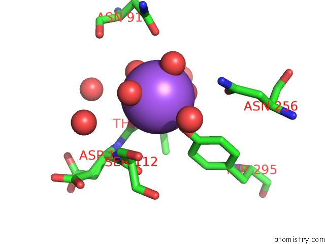
Mono view
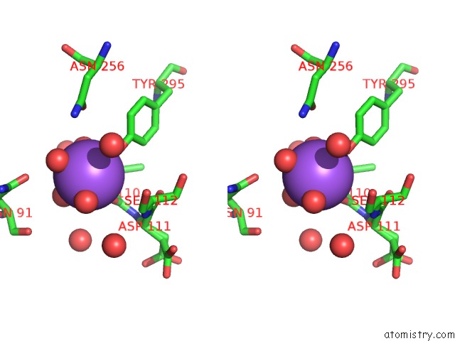
Stereo pair view
Mono view
Stereo pair view
A full contact list of Sodium with other atoms in the Na binding
site number 1 of Glucose/Galactose Binding Protein Bound to 3-O-Methyl D-Glucose within 5.0Å range:
|
Sodium binding site 2 out of 2 in 2qw1
Go back to
Sodium binding site 2 out
of 2 in the Glucose/Galactose Binding Protein Bound to 3-O-Methyl D-Glucose
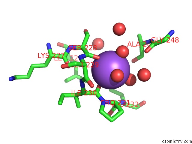
Mono view
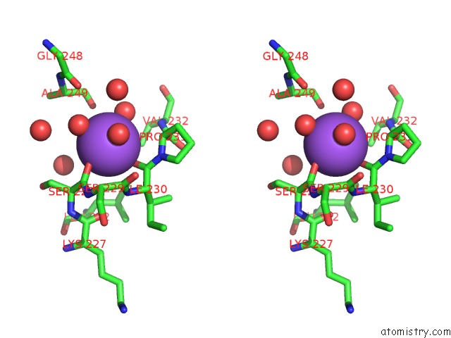
Stereo pair view
Mono view
Stereo pair view
A full contact list of Sodium with other atoms in the Na binding
site number 2 of Glucose/Galactose Binding Protein Bound to 3-O-Methyl D-Glucose within 5.0Å range:
|
Reference:
M.J.Borrok,
Y.Zhu,
K.T.Forest,
L.L.Kiessling.
Structure-Based Design of A Periplasmic Binding Protein Antagonist That Prevents Domain Closure. Acs Chem.Biol. V. 4 447 2009.
ISSN: ISSN 1554-8929
PubMed: 19348466
DOI: 10.1021/CB900021Q
Page generated: Mon Oct 7 04:04:29 2024
ISSN: ISSN 1554-8929
PubMed: 19348466
DOI: 10.1021/CB900021Q
Last articles
F in 7LJEF in 7LTX
F in 7LTP
F in 7LT4
F in 7LRY
F in 7LPX
F in 7LPW
F in 7LQU
F in 7LPF
F in 7LOE