Sodium »
PDB 2qd8-2r22 »
2qr7 »
Sodium in PDB 2qr7: 2.0A X-Ray Structure of C-Terminal Kinase Domain of P90 Ribosomal S6 Kinase 2: Se-Met Derivative
Enzymatic activity of 2.0A X-Ray Structure of C-Terminal Kinase Domain of P90 Ribosomal S6 Kinase 2: Se-Met Derivative
All present enzymatic activity of 2.0A X-Ray Structure of C-Terminal Kinase Domain of P90 Ribosomal S6 Kinase 2: Se-Met Derivative:
2.7.11.1;
2.7.11.1;
Protein crystallography data
The structure of 2.0A X-Ray Structure of C-Terminal Kinase Domain of P90 Ribosomal S6 Kinase 2: Se-Met Derivative, PDB code: 2qr7
was solved by
M.Malakhova,
V.Tereshko,
Z.Dong,
with X-Ray Crystallography technique. A brief refinement statistics is given in the table below:
| Resolution Low / High (Å) | 20.00 / 2.00 |
| Space group | P 41 21 2 |
| Cell size a, b, c (Å), α, β, γ (°) | 46.585, 46.585, 293.990, 90.00, 90.00, 90.00 |
| R / Rfree (%) | 20.3 / 23.8 |
Sodium Binding Sites:
The binding sites of Sodium atom in the 2.0A X-Ray Structure of C-Terminal Kinase Domain of P90 Ribosomal S6 Kinase 2: Se-Met Derivative
(pdb code 2qr7). This binding sites where shown within
5.0 Angstroms radius around Sodium atom.
In total only one binding site of Sodium was determined in the 2.0A X-Ray Structure of C-Terminal Kinase Domain of P90 Ribosomal S6 Kinase 2: Se-Met Derivative, PDB code: 2qr7:
In total only one binding site of Sodium was determined in the 2.0A X-Ray Structure of C-Terminal Kinase Domain of P90 Ribosomal S6 Kinase 2: Se-Met Derivative, PDB code: 2qr7:
Sodium binding site 1 out of 1 in 2qr7
Go back to
Sodium binding site 1 out
of 1 in the 2.0A X-Ray Structure of C-Terminal Kinase Domain of P90 Ribosomal S6 Kinase 2: Se-Met Derivative
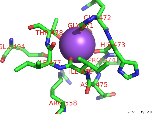
Mono view
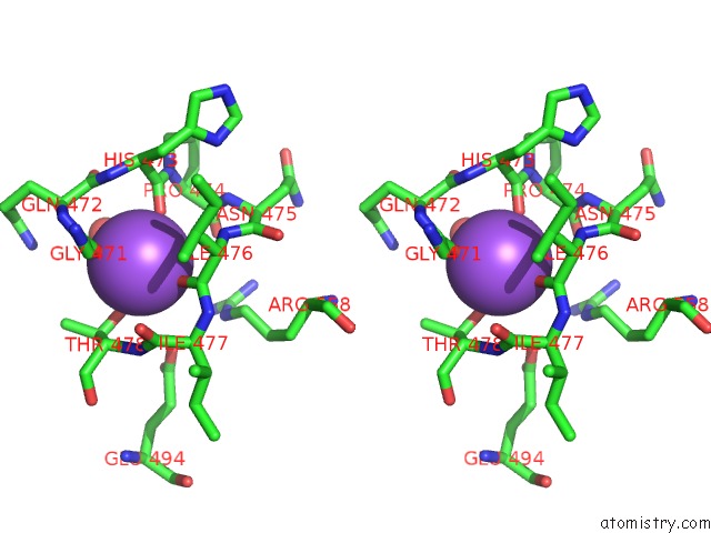
Stereo pair view
Mono view
Stereo pair view
A full contact list of Sodium with other atoms in the Na binding
site number 1 of 2.0A X-Ray Structure of C-Terminal Kinase Domain of P90 Ribosomal S6 Kinase 2: Se-Met Derivative within 5.0Å range:
|
Reference:
M.Malakhova,
V.Tereshko,
S.Y.Lee,
K.Yao,
Y.-Y.Cho,
A.Bode,
Z.Dong.
Structural Basis For Activation of the Autoinhibitory C-Terminal Kinase Domain of P90 RSK2. Nat.Struct.Mol.Biol. V. 15 112 2008.
ISSN: ISSN 1545-9993
PubMed: 18084304
DOI: 10.1038/NSMB1347
Page generated: Mon Oct 7 04:03:46 2024
ISSN: ISSN 1545-9993
PubMed: 18084304
DOI: 10.1038/NSMB1347
Last articles
Zn in 9MJ5Zn in 9HNW
Zn in 9G0L
Zn in 9FNE
Zn in 9DZN
Zn in 9E0I
Zn in 9D32
Zn in 9DAK
Zn in 8ZXC
Zn in 8ZUF