Sodium »
PDB 2fmp-2gg2 »
2g79 »
Sodium in PDB 2g79: Crystal Structure of the R132K:Y134F Mutant of Cellular Retinoic Acid Binding Protein Type II in Complex with All-Trans-Retinal at 1.69 Angstroms Resolution
Protein crystallography data
The structure of Crystal Structure of the R132K:Y134F Mutant of Cellular Retinoic Acid Binding Protein Type II in Complex with All-Trans-Retinal at 1.69 Angstroms Resolution, PDB code: 2g79
was solved by
S.Vaezeslami,
J.H.Geiger,
with X-Ray Crystallography technique. A brief refinement statistics is given in the table below:
| Resolution Low / High (Å) | 39.19 / 1.69 |
| Space group | P 21 21 21 |
| Cell size a, b, c (Å), α, β, γ (°) | 45.493, 46.325, 73.597, 90.00, 90.00, 90.00 |
| R / Rfree (%) | 15.5 / 21.4 |
Sodium Binding Sites:
The binding sites of Sodium atom in the Crystal Structure of the R132K:Y134F Mutant of Cellular Retinoic Acid Binding Protein Type II in Complex with All-Trans-Retinal at 1.69 Angstroms Resolution
(pdb code 2g79). This binding sites where shown within
5.0 Angstroms radius around Sodium atom.
In total 4 binding sites of Sodium where determined in the Crystal Structure of the R132K:Y134F Mutant of Cellular Retinoic Acid Binding Protein Type II in Complex with All-Trans-Retinal at 1.69 Angstroms Resolution, PDB code: 2g79:
Jump to Sodium binding site number: 1; 2; 3; 4;
In total 4 binding sites of Sodium where determined in the Crystal Structure of the R132K:Y134F Mutant of Cellular Retinoic Acid Binding Protein Type II in Complex with All-Trans-Retinal at 1.69 Angstroms Resolution, PDB code: 2g79:
Jump to Sodium binding site number: 1; 2; 3; 4;
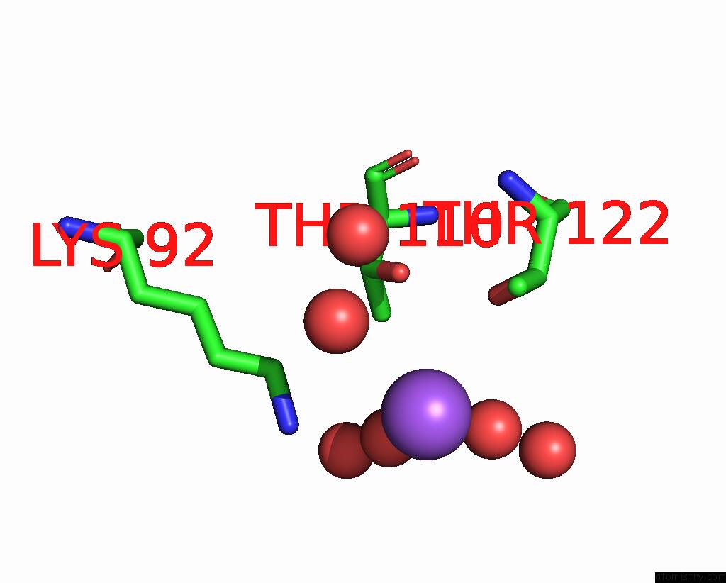
Sodium binding site 1 out of 4 in 2g79
Go back to
Sodium binding site 1 out
of 4 in the Crystal Structure of the R132K:Y134F Mutant of Cellular Retinoic Acid Binding Protein Type II in Complex with All-Trans-Retinal at 1.69 Angstroms Resolution
Mono view

Stereo pair view
Mono view
Stereo pair view
A full contact list of Sodium with other atoms in the Na binding
site number 1 of Crystal Structure of the R132K:Y134F Mutant of Cellular Retinoic Acid Binding Protein Type II in Complex with All-Trans-Retinal at 1.69 Angstroms Resolution within 5.0Å range:
|
Sodium binding site 2 out of 4 in 2g79
Go back to
Sodium binding site 2 out
of 4 in the Crystal Structure of the R132K:Y134F Mutant of Cellular Retinoic Acid Binding Protein Type II in Complex with All-Trans-Retinal at 1.69 Angstroms Resolution

Mono view

Stereo pair view
Mono view
Stereo pair view
A full contact list of Sodium with other atoms in the Na binding
site number 2 of Crystal Structure of the R132K:Y134F Mutant of Cellular Retinoic Acid Binding Protein Type II in Complex with All-Trans-Retinal at 1.69 Angstroms Resolution within 5.0Å range:
|
Sodium binding site 3 out of 4 in 2g79
Go back to
Sodium binding site 3 out
of 4 in the Crystal Structure of the R132K:Y134F Mutant of Cellular Retinoic Acid Binding Protein Type II in Complex with All-Trans-Retinal at 1.69 Angstroms Resolution

Mono view

Stereo pair view
Mono view
Stereo pair view
A full contact list of Sodium with other atoms in the Na binding
site number 3 of Crystal Structure of the R132K:Y134F Mutant of Cellular Retinoic Acid Binding Protein Type II in Complex with All-Trans-Retinal at 1.69 Angstroms Resolution within 5.0Å range:
|
Sodium binding site 4 out of 4 in 2g79
Go back to
Sodium binding site 4 out
of 4 in the Crystal Structure of the R132K:Y134F Mutant of Cellular Retinoic Acid Binding Protein Type II in Complex with All-Trans-Retinal at 1.69 Angstroms Resolution

Mono view

Stereo pair view
Mono view
Stereo pair view
A full contact list of Sodium with other atoms in the Na binding
site number 4 of Crystal Structure of the R132K:Y134F Mutant of Cellular Retinoic Acid Binding Protein Type II in Complex with All-Trans-Retinal at 1.69 Angstroms Resolution within 5.0Å range:
|
Reference:
C.Vasileiou,
S.Vaezeslami,
R.M.Crist,
M.Rabago-Smith,
J.H.Geiger,
B.Borhan.
Protein Design: Reengineering Cellular Retinoic Acid Binding Protein II Into A Rhodopsin Protein Mimic. J.Am.Chem.Soc. V. 129 6140 2007.
ISSN: ISSN 0002-7863
PubMed: 17447762
DOI: 10.1021/JA067546R
Page generated: Mon Oct 7 02:32:36 2024
ISSN: ISSN 0002-7863
PubMed: 17447762
DOI: 10.1021/JA067546R
Last articles
Cl in 5G54Cl in 5G4A
Cl in 5G4Q
Cl in 5G47
Cl in 5G42
Cl in 5G3S
Cl in 5G2P
Cl in 5G2T
Cl in 5G36
Cl in 5G2D