Sodium »
PDB 2fmp-2gg2 »
2fsu »
Sodium in PDB 2fsu: Crystal Structure of the Phnh Protein From Escherichia Coli
Protein crystallography data
The structure of Crystal Structure of the Phnh Protein From Escherichia Coli, PDB code: 2fsu
was solved by
M.A.Adams,
Y.Luo,
D.L.Zechel,
Z.Jia,
Montreal-Kingston Bacterialstructural Genomics Initiative (Bsgi),
with X-Ray Crystallography technique. A brief refinement statistics is given in the table below:
| Resolution Low / High (Å) | 38.92 / 1.70 |
| Space group | C 2 2 21 |
| Cell size a, b, c (Å), α, β, γ (°) | 53.020, 87.418, 75.890, 90.00, 90.00, 90.00 |
| R / Rfree (%) | 18.8 / 24.8 |
Sodium Binding Sites:
The binding sites of Sodium atom in the Crystal Structure of the Phnh Protein From Escherichia Coli
(pdb code 2fsu). This binding sites where shown within
5.0 Angstroms radius around Sodium atom.
In total 4 binding sites of Sodium where determined in the Crystal Structure of the Phnh Protein From Escherichia Coli, PDB code: 2fsu:
Jump to Sodium binding site number: 1; 2; 3; 4;
In total 4 binding sites of Sodium where determined in the Crystal Structure of the Phnh Protein From Escherichia Coli, PDB code: 2fsu:
Jump to Sodium binding site number: 1; 2; 3; 4;
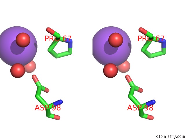
Sodium binding site 1 out of 4 in 2fsu
Go back to
Sodium binding site 1 out
of 4 in the Crystal Structure of the Phnh Protein From Escherichia Coli
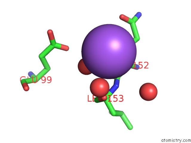
Mono view
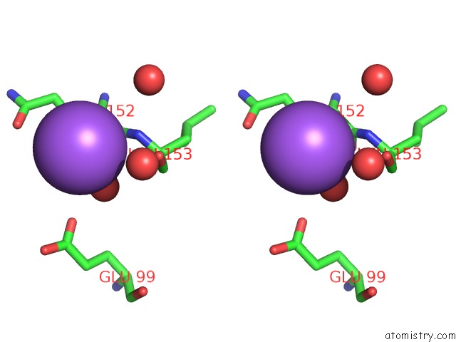
Stereo pair view
Mono view
Stereo pair view
A full contact list of Sodium with other atoms in the Na binding
site number 1 of Crystal Structure of the Phnh Protein From Escherichia Coli within 5.0Å range:
|
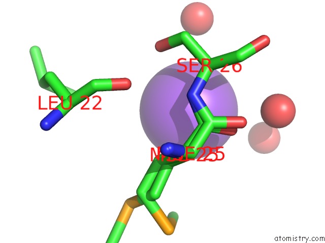
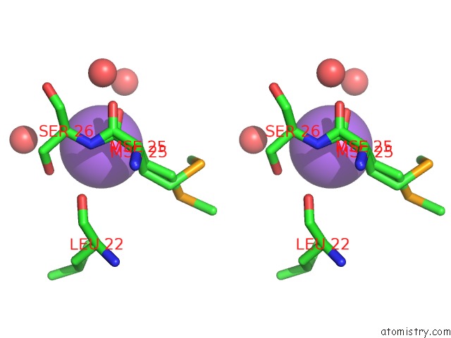
Sodium binding site 2 out of 4 in 2fsu
Go back to
Sodium binding site 2 out
of 4 in the Crystal Structure of the Phnh Protein From Escherichia Coli
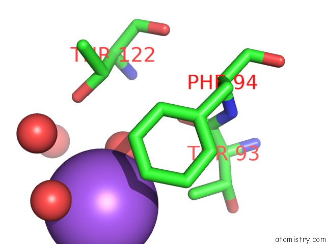
Mono view

Stereo pair view
Mono view
Stereo pair view
A full contact list of Sodium with other atoms in the Na binding
site number 2 of Crystal Structure of the Phnh Protein From Escherichia Coli within 5.0Å range:
|
Sodium binding site 3 out of 4 in 2fsu
Go back to
Sodium binding site 3 out
of 4 in the Crystal Structure of the Phnh Protein From Escherichia Coli
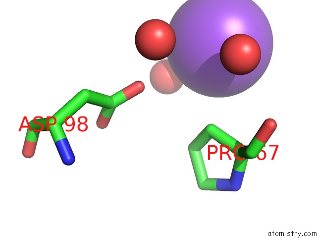
Mono view
Stereo pair view
Mono view
Stereo pair view
A full contact list of Sodium with other atoms in the Na binding
site number 3 of Crystal Structure of the Phnh Protein From Escherichia Coli within 5.0Å range:
|
Sodium binding site 4 out of 4 in 2fsu
Go back to
Sodium binding site 4 out
of 4 in the Crystal Structure of the Phnh Protein From Escherichia Coli
Mono view
Stereo pair view
Mono view
Stereo pair view
A full contact list of Sodium with other atoms in the Na binding
site number 4 of Crystal Structure of the Phnh Protein From Escherichia Coli within 5.0Å range:
|
Reference:
M.A.Adams,
Y.Luo,
B.Hove-Jensen,
S.M.He,
L.M.Van Staalduinen,
D.L.Zechel,
Z.Jia.
Crystal Structure of Phnh: An Essential Component of Carbon-Phosphorus Lyase in Escherichia Coli. J.Bacteriol. V. 190 1072 2008.
ISSN: ISSN 0021-9193
PubMed: 17993513
DOI: 10.1128/JB.01274-07
Page generated: Mon Oct 7 02:29:48 2024
ISSN: ISSN 0021-9193
PubMed: 17993513
DOI: 10.1128/JB.01274-07
Last articles
Zn in 9MJ5Zn in 9HNW
Zn in 9G0L
Zn in 9FNE
Zn in 9DZN
Zn in 9E0I
Zn in 9D32
Zn in 9DAK
Zn in 8ZXC
Zn in 8ZUF