Sodium »
PDB 1x9j-1y4d »
1y0b »
Sodium in PDB 1y0b: Crystal Structure of Xanthine Phosphoribosyltransferase From Bacillus Subtilis.
Protein crystallography data
The structure of Crystal Structure of Xanthine Phosphoribosyltransferase From Bacillus Subtilis., PDB code: 1y0b
was solved by
M.E.Cuff,
R.Wu,
A.Joachimiak,
Midwest Center For Structural Genomics(Mcsg),
with X-Ray Crystallography technique. A brief refinement statistics is given in the table below:
| Resolution Low / High (Å) | 20.00 / 1.80 |
| Space group | P 1 21 1 |
| Cell size a, b, c (Å), α, β, γ (°) | 42.503, 115.902, 73.985, 90.00, 94.30, 90.00 |
| R / Rfree (%) | 17.8 / 22.8 |
Sodium Binding Sites:
The binding sites of Sodium atom in the Crystal Structure of Xanthine Phosphoribosyltransferase From Bacillus Subtilis.
(pdb code 1y0b). This binding sites where shown within
5.0 Angstroms radius around Sodium atom.
In total 8 binding sites of Sodium where determined in the Crystal Structure of Xanthine Phosphoribosyltransferase From Bacillus Subtilis., PDB code: 1y0b:
Jump to Sodium binding site number: 1; 2; 3; 4; 5; 6; 7; 8;
In total 8 binding sites of Sodium where determined in the Crystal Structure of Xanthine Phosphoribosyltransferase From Bacillus Subtilis., PDB code: 1y0b:
Jump to Sodium binding site number: 1; 2; 3; 4; 5; 6; 7; 8;
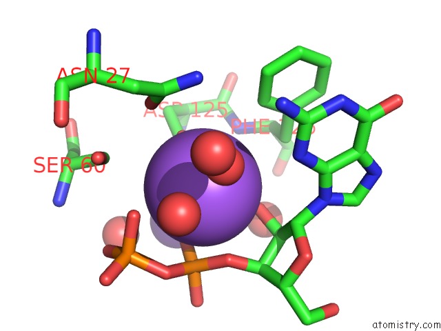
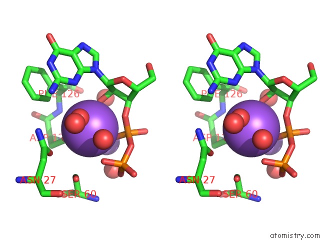
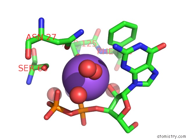
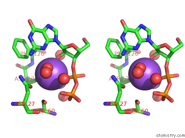
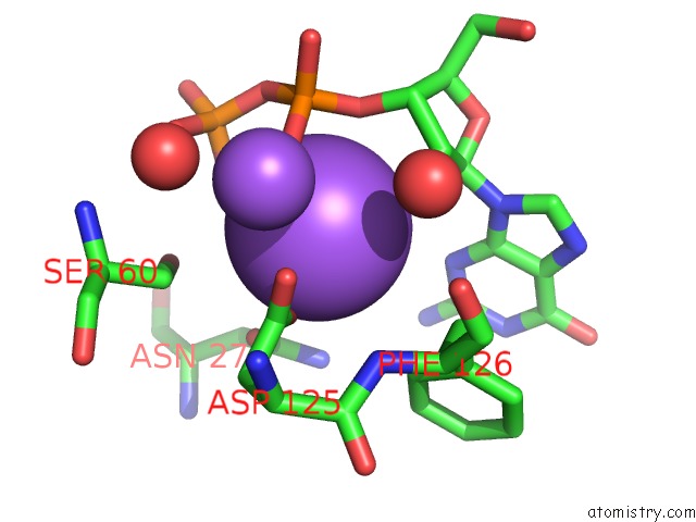
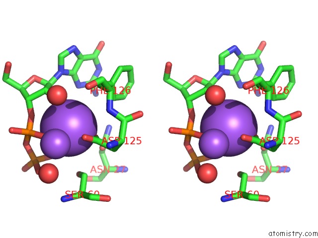
Sodium binding site 1 out of 8 in 1y0b
Go back to
Sodium binding site 1 out
of 8 in the Crystal Structure of Xanthine Phosphoribosyltransferase From Bacillus Subtilis.
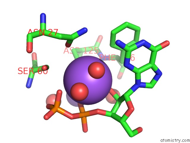
Mono view
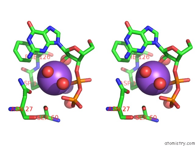
Stereo pair view
Mono view
Stereo pair view
A full contact list of Sodium with other atoms in the Na binding
site number 1 of Crystal Structure of Xanthine Phosphoribosyltransferase From Bacillus Subtilis. within 5.0Å range:
|
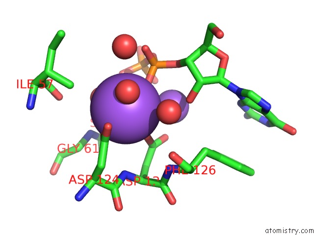
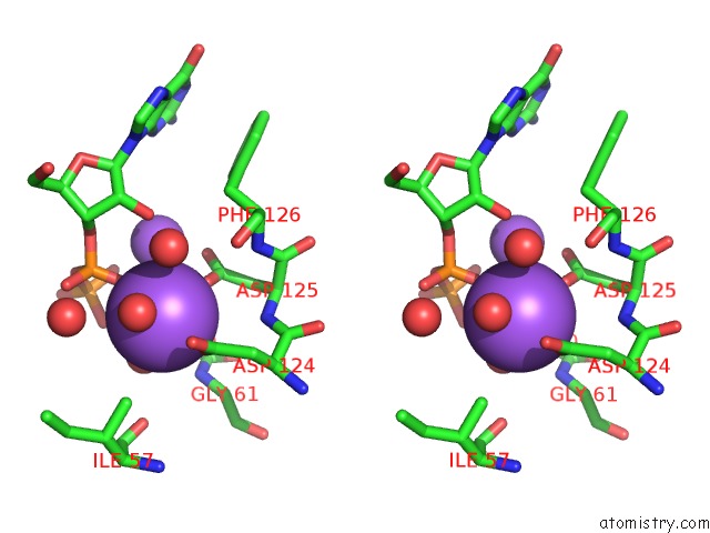
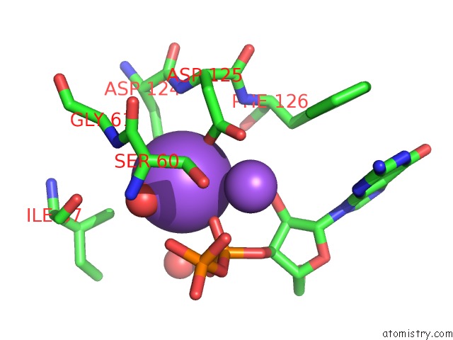
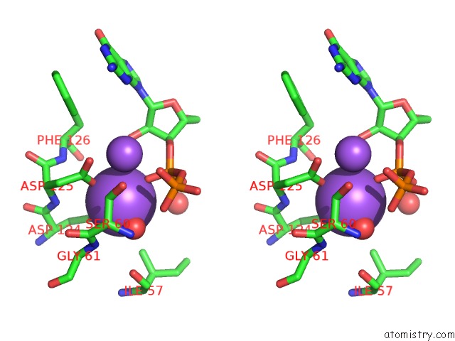
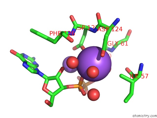
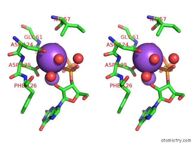
Sodium binding site 2 out of 8 in 1y0b
Go back to
Sodium binding site 2 out
of 8 in the Crystal Structure of Xanthine Phosphoribosyltransferase From Bacillus Subtilis.
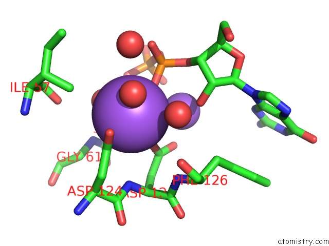
Mono view
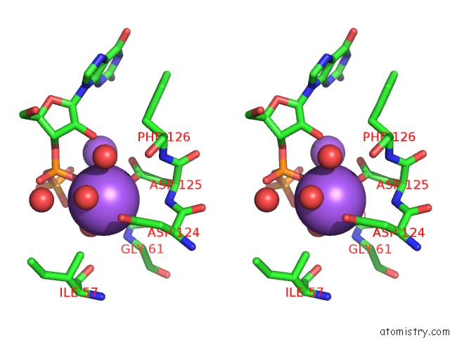
Stereo pair view
Mono view
Stereo pair view
A full contact list of Sodium with other atoms in the Na binding
site number 2 of Crystal Structure of Xanthine Phosphoribosyltransferase From Bacillus Subtilis. within 5.0Å range:
|
Sodium binding site 3 out of 8 in 1y0b
Go back to
Sodium binding site 3 out
of 8 in the Crystal Structure of Xanthine Phosphoribosyltransferase From Bacillus Subtilis.
Mono view
Stereo pair view
Mono view
Stereo pair view
A full contact list of Sodium with other atoms in the Na binding
site number 3 of Crystal Structure of Xanthine Phosphoribosyltransferase From Bacillus Subtilis. within 5.0Å range:
|
Sodium binding site 4 out of 8 in 1y0b
Go back to
Sodium binding site 4 out
of 8 in the Crystal Structure of Xanthine Phosphoribosyltransferase From Bacillus Subtilis.
Mono view
Stereo pair view
Mono view
Stereo pair view
A full contact list of Sodium with other atoms in the Na binding
site number 4 of Crystal Structure of Xanthine Phosphoribosyltransferase From Bacillus Subtilis. within 5.0Å range:
|
Sodium binding site 5 out of 8 in 1y0b
Go back to
Sodium binding site 5 out
of 8 in the Crystal Structure of Xanthine Phosphoribosyltransferase From Bacillus Subtilis.
Mono view
Stereo pair view
Mono view
Stereo pair view
A full contact list of Sodium with other atoms in the Na binding
site number 5 of Crystal Structure of Xanthine Phosphoribosyltransferase From Bacillus Subtilis. within 5.0Å range:
|
Sodium binding site 6 out of 8 in 1y0b
Go back to
Sodium binding site 6 out
of 8 in the Crystal Structure of Xanthine Phosphoribosyltransferase From Bacillus Subtilis.
Mono view
Stereo pair view
Mono view
Stereo pair view
A full contact list of Sodium with other atoms in the Na binding
site number 6 of Crystal Structure of Xanthine Phosphoribosyltransferase From Bacillus Subtilis. within 5.0Å range:
|
Sodium binding site 7 out of 8 in 1y0b
Go back to
Sodium binding site 7 out
of 8 in the Crystal Structure of Xanthine Phosphoribosyltransferase From Bacillus Subtilis.
Mono view
Stereo pair view
Mono view
Stereo pair view
A full contact list of Sodium with other atoms in the Na binding
site number 7 of Crystal Structure of Xanthine Phosphoribosyltransferase From Bacillus Subtilis. within 5.0Å range:
|
Sodium binding site 8 out of 8 in 1y0b
Go back to
Sodium binding site 8 out
of 8 in the Crystal Structure of Xanthine Phosphoribosyltransferase From Bacillus Subtilis.
Mono view
Stereo pair view
Mono view
Stereo pair view
A full contact list of Sodium with other atoms in the Na binding
site number 8 of Crystal Structure of Xanthine Phosphoribosyltransferase From Bacillus Subtilis. within 5.0Å range:
|
Reference:
M.E.Cuff,
R.Wu,
A.Joachimiak,
Mcsg.
Crystal Structure of Xanthine Phosphoribosyltransferase From Bacillus Subtilis To Be Published.
Page generated: Mon Oct 7 00:35:32 2024
Last articles
Fe in 2YXOFe in 2YRS
Fe in 2YXC
Fe in 2YNM
Fe in 2YVJ
Fe in 2YP1
Fe in 2YU2
Fe in 2YU1
Fe in 2YQB
Fe in 2YOO