Sodium »
PDB 1x9j-1y4d »
1xnz »
Sodium in PDB 1xnz: Crystal Structure of Mn(II) Form of E. Coli. Methionine Aminopeptidase in Complex with 5-(2-Chlorophenyl)Furan-2- Carboxylic Acid
Enzymatic activity of Crystal Structure of Mn(II) Form of E. Coli. Methionine Aminopeptidase in Complex with 5-(2-Chlorophenyl)Furan-2- Carboxylic Acid
All present enzymatic activity of Crystal Structure of Mn(II) Form of E. Coli. Methionine Aminopeptidase in Complex with 5-(2-Chlorophenyl)Furan-2- Carboxylic Acid:
3.4.11.18;
3.4.11.18;
Protein crystallography data
The structure of Crystal Structure of Mn(II) Form of E. Coli. Methionine Aminopeptidase in Complex with 5-(2-Chlorophenyl)Furan-2- Carboxylic Acid, PDB code: 1xnz
was solved by
Q.-Z.Ye,
S.-X.Xie,
M.Huang,
W.-J.Huang,
J.-P.Lu,
Z.-Q.Ma,
with X-Ray Crystallography technique. A brief refinement statistics is given in the table below:
| Resolution Low / High (Å) | 10.00 / 1.52 |
| Space group | P 1 21 1 |
| Cell size a, b, c (Å), α, β, γ (°) | 37.600, 60.300, 49.700, 90.00, 104.80, 90.00 |
| R / Rfree (%) | 21.9 / 25 |
Other elements in 1xnz:
The structure of Crystal Structure of Mn(II) Form of E. Coli. Methionine Aminopeptidase in Complex with 5-(2-Chlorophenyl)Furan-2- Carboxylic Acid also contains other interesting chemical elements:
| Manganese | (Mn) | 2 atoms |
| Chlorine | (Cl) | 1 atom |
Sodium Binding Sites:
The binding sites of Sodium atom in the Crystal Structure of Mn(II) Form of E. Coli. Methionine Aminopeptidase in Complex with 5-(2-Chlorophenyl)Furan-2- Carboxylic Acid
(pdb code 1xnz). This binding sites where shown within
5.0 Angstroms radius around Sodium atom.
In total only one binding site of Sodium was determined in the Crystal Structure of Mn(II) Form of E. Coli. Methionine Aminopeptidase in Complex with 5-(2-Chlorophenyl)Furan-2- Carboxylic Acid, PDB code: 1xnz:
In total only one binding site of Sodium was determined in the Crystal Structure of Mn(II) Form of E. Coli. Methionine Aminopeptidase in Complex with 5-(2-Chlorophenyl)Furan-2- Carboxylic Acid, PDB code: 1xnz:
Sodium binding site 1 out of 1 in 1xnz
Go back to
Sodium binding site 1 out
of 1 in the Crystal Structure of Mn(II) Form of E. Coli. Methionine Aminopeptidase in Complex with 5-(2-Chlorophenyl)Furan-2- Carboxylic Acid
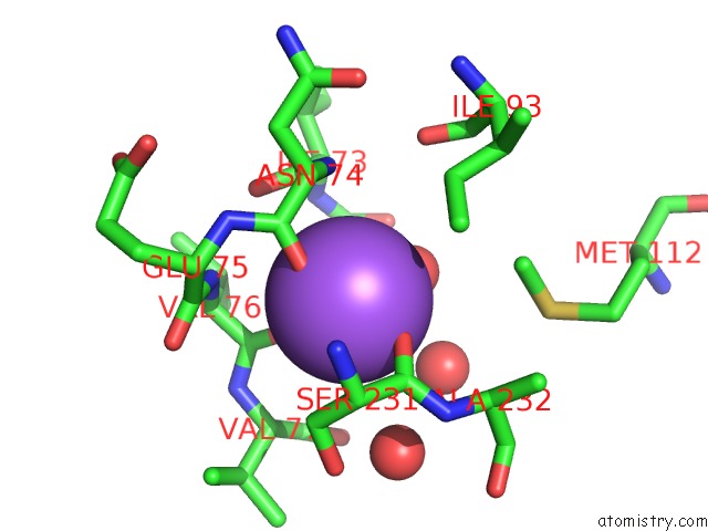
Mono view
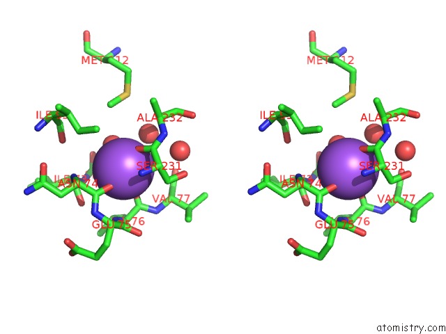
Stereo pair view
Mono view
Stereo pair view
A full contact list of Sodium with other atoms in the Na binding
site number 1 of Crystal Structure of Mn(II) Form of E. Coli. Methionine Aminopeptidase in Complex with 5-(2-Chlorophenyl)Furan-2- Carboxylic Acid within 5.0Å range:
|
Reference:
Q.-Z.Ye,
S.-X.Xie,
M.Huang,
W.-J.Huang,
J.-P.Lu,
Z.-Q.Ma.
Metalloform-Selective Inhibitors of Escherichia Coli Methionine Aminopeptidase and X-Ray Structure of A Mn(II)-Form Enzyme Complexed with An Inhibitor. J.Am.Chem.Soc. V. 126 13940 2004.
ISSN: ISSN 0002-7863
PubMed: 15506752
DOI: 10.1021/JA045864P
Page generated: Mon Oct 7 00:33:36 2024
ISSN: ISSN 0002-7863
PubMed: 15506752
DOI: 10.1021/JA045864P
Last articles
Zn in 9J0NZn in 9J0O
Zn in 9J0P
Zn in 9FJX
Zn in 9EKB
Zn in 9C0F
Zn in 9CAH
Zn in 9CH0
Zn in 9CH3
Zn in 9CH1