Sodium »
PDB 1x9j-1y4d »
1xdf »
Sodium in PDB 1xdf: Crystal Structure of Pathogenesis-Related Protein Llpr-10.2A From Yellow Lupine
Protein crystallography data
The structure of Crystal Structure of Pathogenesis-Related Protein Llpr-10.2A From Yellow Lupine, PDB code: 1xdf
was solved by
O.Pasternak,
J.Biesiadka,
R.Dolot,
L.Handschuh,
G.Bujacz,
M.M.Sikorski,
M.Jaskolski,
with X-Ray Crystallography technique. A brief refinement statistics is given in the table below:
| Resolution Low / High (Å) | 20.00 / 1.90 |
| Space group | P 21 21 21 |
| Cell size a, b, c (Å), α, β, γ (°) | 48.956, 69.243, 112.918, 90.00, 90.00, 90.00 |
| R / Rfree (%) | 20.5 / 25.1 |
Sodium Binding Sites:
The binding sites of Sodium atom in the Crystal Structure of Pathogenesis-Related Protein Llpr-10.2A From Yellow Lupine
(pdb code 1xdf). This binding sites where shown within
5.0 Angstroms radius around Sodium atom.
In total only one binding site of Sodium was determined in the Crystal Structure of Pathogenesis-Related Protein Llpr-10.2A From Yellow Lupine, PDB code: 1xdf:
In total only one binding site of Sodium was determined in the Crystal Structure of Pathogenesis-Related Protein Llpr-10.2A From Yellow Lupine, PDB code: 1xdf:
Sodium binding site 1 out of 1 in 1xdf
Go back to
Sodium binding site 1 out
of 1 in the Crystal Structure of Pathogenesis-Related Protein Llpr-10.2A From Yellow Lupine
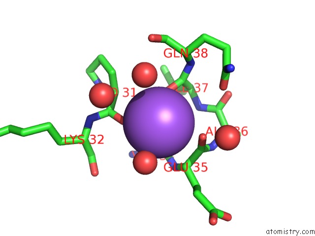
Mono view
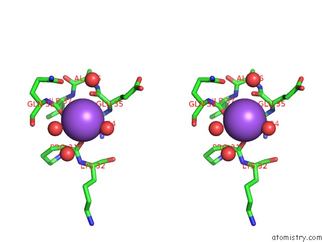
Stereo pair view
Mono view
Stereo pair view
A full contact list of Sodium with other atoms in the Na binding
site number 1 of Crystal Structure of Pathogenesis-Related Protein Llpr-10.2A From Yellow Lupine within 5.0Å range:
|
Reference:
O.Pasternak,
J.Biesiadka,
R.Dolot,
L.Handschuh,
G.Bujacz,
M.M.Sikorski,
M.Jaskolski.
Structure of A Yellow Lupin Pathogenesis-Related Pr-10 Protein Belonging to A Novel Subclass. Acta Crystallogr.,Sect.D V. 61 99 2005.
ISSN: ISSN 0907-4449
PubMed: 15608381
DOI: 10.1107/S0907444904028173
Page generated: Mon Oct 7 00:31:50 2024
ISSN: ISSN 0907-4449
PubMed: 15608381
DOI: 10.1107/S0907444904028173
Last articles
Zn in 9J0NZn in 9J0O
Zn in 9J0P
Zn in 9FJX
Zn in 9EKB
Zn in 9C0F
Zn in 9CAH
Zn in 9CH0
Zn in 9CH3
Zn in 9CH1