Sodium »
PDB 1nnh-1oan »
1o68 »
Sodium in PDB 1o68: Crystal Structure of 3-Methyl-2-Oxobutanoate Hydroxymethyltransferase
Enzymatic activity of Crystal Structure of 3-Methyl-2-Oxobutanoate Hydroxymethyltransferase
All present enzymatic activity of Crystal Structure of 3-Methyl-2-Oxobutanoate Hydroxymethyltransferase:
2.1.2.11;
2.1.2.11;
Protein crystallography data
The structure of Crystal Structure of 3-Methyl-2-Oxobutanoate Hydroxymethyltransferase, PDB code: 1o68
was solved by
Structural Genomix,
with X-Ray Crystallography technique. A brief refinement statistics is given in the table below:
| Resolution Low / High (Å) | 69.00 / 2.10 |
| Space group | C 1 2 1 |
| Cell size a, b, c (Å), α, β, γ (°) | 164.419, 111.238, 98.807, 90.00, 122.93, 90.00 |
| R / Rfree (%) | 18.6 / 22.4 |
Sodium Binding Sites:
The binding sites of Sodium atom in the Crystal Structure of 3-Methyl-2-Oxobutanoate Hydroxymethyltransferase
(pdb code 1o68). This binding sites where shown within
5.0 Angstroms radius around Sodium atom.
In total 4 binding sites of Sodium where determined in the Crystal Structure of 3-Methyl-2-Oxobutanoate Hydroxymethyltransferase, PDB code: 1o68:
Jump to Sodium binding site number: 1; 2; 3; 4;
In total 4 binding sites of Sodium where determined in the Crystal Structure of 3-Methyl-2-Oxobutanoate Hydroxymethyltransferase, PDB code: 1o68:
Jump to Sodium binding site number: 1; 2; 3; 4;
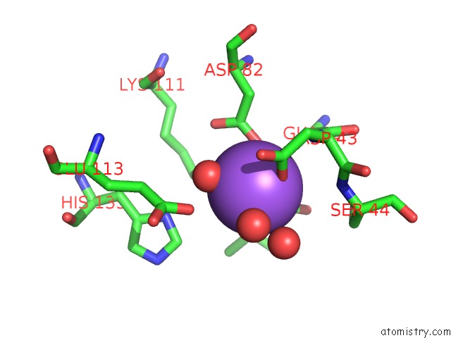
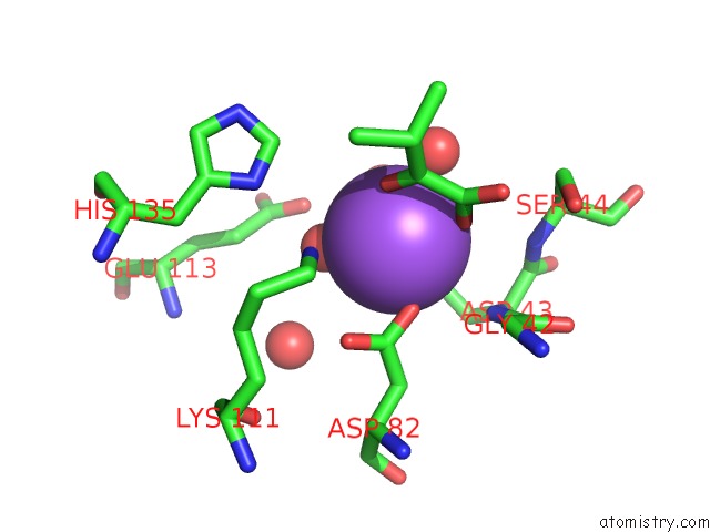
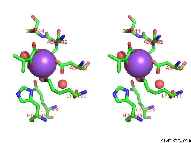
Sodium binding site 1 out of 4 in 1o68
Go back to
Sodium binding site 1 out
of 4 in the Crystal Structure of 3-Methyl-2-Oxobutanoate Hydroxymethyltransferase
Mono view
Stereo pair view
Mono view
Stereo pair view
A full contact list of Sodium with other atoms in the Na binding
site number 1 of Crystal Structure of 3-Methyl-2-Oxobutanoate Hydroxymethyltransferase within 5.0Å range:
|
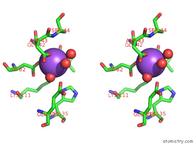
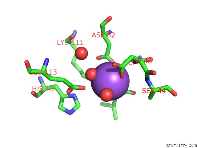
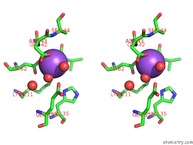
Sodium binding site 2 out of 4 in 1o68
Go back to
Sodium binding site 2 out
of 4 in the Crystal Structure of 3-Methyl-2-Oxobutanoate Hydroxymethyltransferase
Mono view
Stereo pair view
Mono view
Stereo pair view
A full contact list of Sodium with other atoms in the Na binding
site number 2 of Crystal Structure of 3-Methyl-2-Oxobutanoate Hydroxymethyltransferase within 5.0Å range:
|
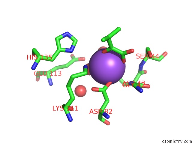
Sodium binding site 3 out of 4 in 1o68
Go back to
Sodium binding site 3 out
of 4 in the Crystal Structure of 3-Methyl-2-Oxobutanoate Hydroxymethyltransferase
Mono view
Stereo pair view
Mono view
Stereo pair view
A full contact list of Sodium with other atoms in the Na binding
site number 3 of Crystal Structure of 3-Methyl-2-Oxobutanoate Hydroxymethyltransferase within 5.0Å range:
|
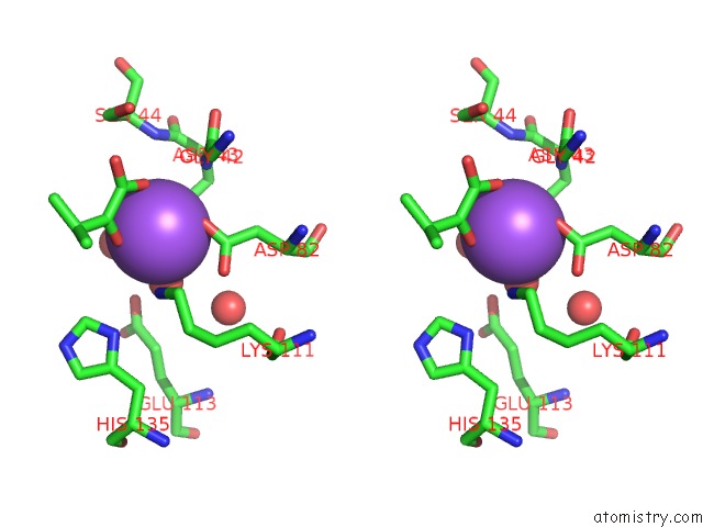
Sodium binding site 4 out of 4 in 1o68
Go back to
Sodium binding site 4 out
of 4 in the Crystal Structure of 3-Methyl-2-Oxobutanoate Hydroxymethyltransferase
Mono view
Stereo pair view
Mono view
Stereo pair view
A full contact list of Sodium with other atoms in the Na binding
site number 4 of Crystal Structure of 3-Methyl-2-Oxobutanoate Hydroxymethyltransferase within 5.0Å range:
|
Reference:
J.Badger,
J.M.Sauder,
J.M.Adams,
S.Antonysamy,
K.Bain,
M.G.Bergseid,
S.G.Buchanan,
M.D.Buchanan,
Y.Batiyenko,
J.A.Christopher,
S.Emtage,
A.Eroshkina,
I.Feil,
E.B.Furlong,
K.S.Gajiwala,
X.Gao,
D.He,
J.Hendle,
A.Huber,
K.Hoda,
P.Kearins,
C.Kissinger,
B.Laubert,
H.A.Lewis,
J.Lin,
K.Loomis,
D.Lorimer,
G.Louie,
M.Maletic,
C.D.Marsh,
I.Miller,
J.Molinari,
H.J.Muller-Dieckmann,
J.M.Newman,
B.W.Noland,
B.Pagarigan,
F.Park,
T.S.Peat,
K.W.Post,
S.Radojicic,
A.Ramos,
R.Romero,
M.E.Rutter,
W.E.Sanderson,
K.D.Schwinn,
J.Tresser,
J.Winhoven,
T.A.Wright,
L.Wu,
J.Xu,
T.J.Harris.
Structural Analysis of A Set of Proteins Resulting From A Bacterial Genomics Project Proteins V. 60 787 2005.
ISSN: ISSN 0887-3585
PubMed: 16021622
DOI: 10.1002/PROT.20541
Page generated: Sun Aug 17 06:50:44 2025
ISSN: ISSN 0887-3585
PubMed: 16021622
DOI: 10.1002/PROT.20541
Last articles
Na in 3IANNa in 3I8T
Na in 3I78
Na in 3I3E
Na in 3I6V
Na in 3I6O
Na in 3I6E
Na in 3I3D
Na in 3I4Q
Na in 3I44