Sodium »
PDB 1ghy-1hn1 »
1hn0 »
Sodium in PDB 1hn0: Crystal Structure of Chondroitin Abc Lyase I From Proteus Vulgaris at 1.9 Angstroms Resolution
Enzymatic activity of Crystal Structure of Chondroitin Abc Lyase I From Proteus Vulgaris at 1.9 Angstroms Resolution
All present enzymatic activity of Crystal Structure of Chondroitin Abc Lyase I From Proteus Vulgaris at 1.9 Angstroms Resolution:
4.2.2.4;
4.2.2.4;
Protein crystallography data
The structure of Crystal Structure of Chondroitin Abc Lyase I From Proteus Vulgaris at 1.9 Angstroms Resolution, PDB code: 1hn0
was solved by
W.Huang,
M.Cygler,
with X-Ray Crystallography technique. A brief refinement statistics is given in the table below:
| Resolution Low / High (Å) | 19.88 / 1.90 |
| Space group | P 21 21 21 |
| Cell size a, b, c (Å), α, β, γ (°) | 49.280, 95.140, 229.950, 90.00, 90.00, 90.00 |
| R / Rfree (%) | 15.6 / 21.2 |
Sodium Binding Sites:
The binding sites of Sodium atom in the Crystal Structure of Chondroitin Abc Lyase I From Proteus Vulgaris at 1.9 Angstroms Resolution
(pdb code 1hn0). This binding sites where shown within
5.0 Angstroms radius around Sodium atom.
In total only one binding site of Sodium was determined in the Crystal Structure of Chondroitin Abc Lyase I From Proteus Vulgaris at 1.9 Angstroms Resolution, PDB code: 1hn0:
In total only one binding site of Sodium was determined in the Crystal Structure of Chondroitin Abc Lyase I From Proteus Vulgaris at 1.9 Angstroms Resolution, PDB code: 1hn0:
Sodium binding site 1 out of 1 in 1hn0
Go back to
Sodium binding site 1 out
of 1 in the Crystal Structure of Chondroitin Abc Lyase I From Proteus Vulgaris at 1.9 Angstroms Resolution
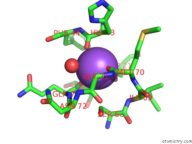
Mono view
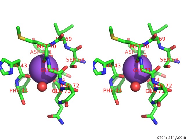
Stereo pair view
Mono view
Stereo pair view
A full contact list of Sodium with other atoms in the Na binding
site number 1 of Crystal Structure of Chondroitin Abc Lyase I From Proteus Vulgaris at 1.9 Angstroms Resolution within 5.0Å range:
|
Reference:
W.Huang,
V.V.Lunin,
Y.Li,
S.Suzuki,
N.Sugiura,
H.Miyazono,
M.Cygler.
Crystal Structure of Proteus Vulgaris Chondroitin Sulfate Abc Lyase I at 1.9 Angstroms Resolution J.Mol.Biol. V. 328 623 2003.
ISSN: ISSN 0022-2836
PubMed: 12706721
DOI: 10.1016/S0022-2836(03)00345-0
Page generated: Sun Aug 17 05:13:49 2025
ISSN: ISSN 0022-2836
PubMed: 12706721
DOI: 10.1016/S0022-2836(03)00345-0
Last articles
Na in 8WCINa in 8WG6
Na in 8WFW
Na in 8WFV
Na in 8WFK
Na in 8WFI
Na in 8WEN
Na in 8WCH
Na in 8WC0
Na in 8W9N