Sodium »
PDB 1ghy-1hn1 »
1gw1 »
Sodium in PDB 1gw1: Substrate Distortion By Beta-Mannanase From Pseudomonas Cellulosa
Enzymatic activity of Substrate Distortion By Beta-Mannanase From Pseudomonas Cellulosa
All present enzymatic activity of Substrate Distortion By Beta-Mannanase From Pseudomonas Cellulosa:
3.2.1.78;
3.2.1.78;
Protein crystallography data
The structure of Substrate Distortion By Beta-Mannanase From Pseudomonas Cellulosa, PDB code: 1gw1
was solved by
V.Ducros,
D.L.Zechel,
H.J.Gilbert,
L.Szabo,
S.G.Withers,
G.J.Davies,
with X-Ray Crystallography technique. A brief refinement statistics is given in the table below:
| Resolution Low / High (Å) | 20.00 / 1.65 |
| Space group | P 41 |
| Cell size a, b, c (Å), α, β, γ (°) | 93.488, 93.488, 53.719, 90.00, 90.00, 90.00 |
| R / Rfree (%) | 13.8 / 16.7 |
Other elements in 1gw1:
The structure of Substrate Distortion By Beta-Mannanase From Pseudomonas Cellulosa also contains other interesting chemical elements:
| Fluorine | (F) | 2 atoms |
| Zinc | (Zn) | 2 atoms |
Sodium Binding Sites:
The binding sites of Sodium atom in the Substrate Distortion By Beta-Mannanase From Pseudomonas Cellulosa
(pdb code 1gw1). This binding sites where shown within
5.0 Angstroms radius around Sodium atom.
In total only one binding site of Sodium was determined in the Substrate Distortion By Beta-Mannanase From Pseudomonas Cellulosa, PDB code: 1gw1:
In total only one binding site of Sodium was determined in the Substrate Distortion By Beta-Mannanase From Pseudomonas Cellulosa, PDB code: 1gw1:
Sodium binding site 1 out of 1 in 1gw1
Go back to
Sodium binding site 1 out
of 1 in the Substrate Distortion By Beta-Mannanase From Pseudomonas Cellulosa
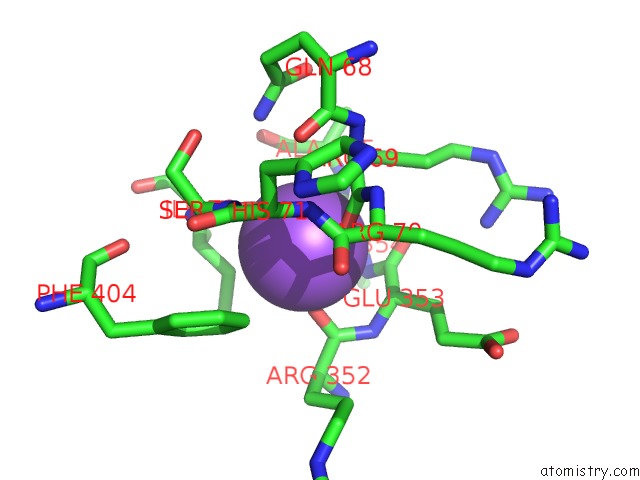
Mono view
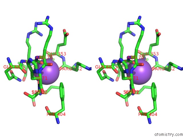
Stereo pair view
Mono view
Stereo pair view
A full contact list of Sodium with other atoms in the Na binding
site number 1 of Substrate Distortion By Beta-Mannanase From Pseudomonas Cellulosa within 5.0Å range:
|
Reference:
V.Ducros,
D.L.Zechel,
G.Murshudov,
H.J.Gilbert,
L.Szabo,
D.Stoll,
S.G.Withers,
G.J.Davies.
Substrate Distortion By A Beta-Mannanase: Snapshots of the Michaelis and Covalent-Intermediate Complexes Suggest A B2,5 Conformation For the Transition State Angew.Chem.Int.Ed.Engl. V. 41 2824 2002.
ISSN: ISSN 1433-7851
PubMed: 12203498
DOI: 10.1002/1521-3773(20020802)41:15<2824::AID-ANIE2824>3.0.CO;2
Page generated: Sun Oct 6 18:38:34 2024
ISSN: ISSN 1433-7851
PubMed: 12203498
DOI: 10.1002/1521-3773(20020802)41:15<2824::AID-ANIE2824>3.0.CO;2
Last articles
Zn in 9MJ5Zn in 9HNW
Zn in 9G0L
Zn in 9FNE
Zn in 9DZN
Zn in 9E0I
Zn in 9D32
Zn in 9DAK
Zn in 8ZXC
Zn in 8ZUF