Sodium »
PDB 1ghy-1hn1 »
1gv5 »
Sodium in PDB 1gv5: Crystal Structure of C-Myb R2
Protein crystallography data
The structure of Crystal Structure of C-Myb R2, PDB code: 1gv5
was solved by
T.H.Tahirov,
K.Ogata,
with X-Ray Crystallography technique. A brief refinement statistics is given in the table below:
| Resolution Low / High (Å) | 14.26 / 1.58 |
| Space group | P 21 21 21 |
| Cell size a, b, c (Å), α, β, γ (°) | 28.832, 40.182, 49.229, 90.00, 90.00, 90.00 |
| R / Rfree (%) | 17.8 / 19 |
Sodium Binding Sites:
The binding sites of Sodium atom in the Crystal Structure of C-Myb R2
(pdb code 1gv5). This binding sites where shown within
5.0 Angstroms radius around Sodium atom.
In total only one binding site of Sodium was determined in the Crystal Structure of C-Myb R2, PDB code: 1gv5:
In total only one binding site of Sodium was determined in the Crystal Structure of C-Myb R2, PDB code: 1gv5:
Sodium binding site 1 out of 1 in 1gv5
Go back to
Sodium binding site 1 out
of 1 in the Crystal Structure of C-Myb R2
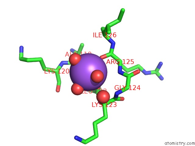
Mono view
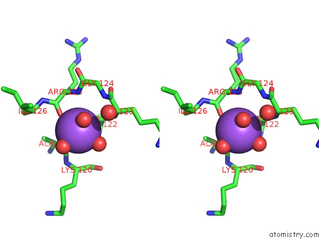
Stereo pair view
Mono view
Stereo pair view
A full contact list of Sodium with other atoms in the Na binding
site number 1 of Crystal Structure of C-Myb R2 within 5.0Å range:
|
Reference:
T.H.Tahirov,
H.Morii,
H.Uedaira,
M.Sasaki,
A.Sarai,
S.Adachi,
S.Y.Park,
N.Kamiya,
K.Ogata.
Crystal Structure of C-Myb Dna-Binding Domain: Specific Na+ Binding and Correlation with uc(Nmr) Structure To Be Published.
Page generated: Sun Oct 6 18:38:03 2024
Last articles
Cl in 5SIOCl in 5SID
Cl in 5SIE
Cl in 5SIL
Cl in 5SHT
Cl in 5SIF
Cl in 5SHY
Cl in 5SHL
Cl in 5SHM
Cl in 5SHG