Sodium »
PDB 1ghy-1hn1 »
1goh »
Sodium in PDB 1goh: Novel Thioether Bond Revealed By A 1.7 Angstroms Crystal Structure of Galactose Oxidase
Enzymatic activity of Novel Thioether Bond Revealed By A 1.7 Angstroms Crystal Structure of Galactose Oxidase
All present enzymatic activity of Novel Thioether Bond Revealed By A 1.7 Angstroms Crystal Structure of Galactose Oxidase:
1.1.3.9;
1.1.3.9;
Protein crystallography data
The structure of Novel Thioether Bond Revealed By A 1.7 Angstroms Crystal Structure of Galactose Oxidase, PDB code: 1goh
was solved by
N.Ito,
S.E.V.Phillips,
P.F.Knowles,
with X-Ray Crystallography technique. A brief refinement statistics is given in the table below:
| Resolution Low / High (Å) | 10.00 / 2.20 |
| Space group | C 1 2 1 |
| Cell size a, b, c (Å), α, β, γ (°) | 98.000, 89.400, 86.700, 90.00, 117.80, 90.00 |
| R / Rfree (%) | n/a / n/a |
Sodium Binding Sites:
The binding sites of Sodium atom in the Novel Thioether Bond Revealed By A 1.7 Angstroms Crystal Structure of Galactose Oxidase
(pdb code 1goh). This binding sites where shown within
5.0 Angstroms radius around Sodium atom.
In total only one binding site of Sodium was determined in the Novel Thioether Bond Revealed By A 1.7 Angstroms Crystal Structure of Galactose Oxidase, PDB code: 1goh:
In total only one binding site of Sodium was determined in the Novel Thioether Bond Revealed By A 1.7 Angstroms Crystal Structure of Galactose Oxidase, PDB code: 1goh:
Sodium binding site 1 out of 1 in 1goh
Go back to
Sodium binding site 1 out
of 1 in the Novel Thioether Bond Revealed By A 1.7 Angstroms Crystal Structure of Galactose Oxidase
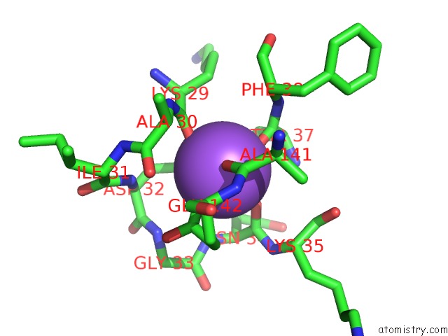
Mono view
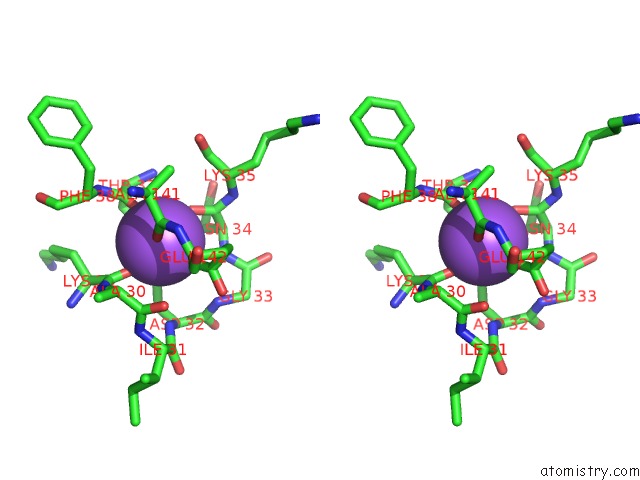
Stereo pair view
Mono view
Stereo pair view
A full contact list of Sodium with other atoms in the Na binding
site number 1 of Novel Thioether Bond Revealed By A 1.7 Angstroms Crystal Structure of Galactose Oxidase within 5.0Å range:
|
Reference:
N.Ito,
S.E.Phillips,
C.Stevens,
Z.B.Ogel,
M.J.Mcpherson,
J.N.Keen,
K.D.Yadav,
P.F.Knowles.
Novel Thioether Bond Revealed By A 1.7 A Crystal Structure of Galactose Oxidase. Nature V. 350 87 1991.
ISSN: ISSN 0028-0836
PubMed: 2002850
DOI: 10.1038/350087A0
Page generated: Sun Oct 6 18:37:43 2024
ISSN: ISSN 0028-0836
PubMed: 2002850
DOI: 10.1038/350087A0
Last articles
Zn in 9MJ5Zn in 9HNW
Zn in 9G0L
Zn in 9FNE
Zn in 9DZN
Zn in 9E0I
Zn in 9D32
Zn in 9DAK
Zn in 8ZXC
Zn in 8ZUF