Sodium »
PDB 7vmf-7wys »
7w52 »
Sodium in PDB 7w52: Crystal Structure of Fragmin Domain-1 (15-160) in Complex with Actin
Protein crystallography data
The structure of Crystal Structure of Fragmin Domain-1 (15-160) in Complex with Actin, PDB code: 7w52
was solved by
S.Takeda,
with X-Ray Crystallography technique. A brief refinement statistics is given in the table below:
| Resolution Low / High (Å) | 46.67 / 2.00 |
| Space group | P 21 21 21 |
| Cell size a, b, c (Å), α, β, γ (°) | 56.987, 96.26, 426.923, 90, 90, 90 |
| R / Rfree (%) | 21 / 24.6 |
Other elements in 7w52:
The structure of Crystal Structure of Fragmin Domain-1 (15-160) in Complex with Actin also contains other interesting chemical elements:
| Calcium | (Ca) | 12 atoms |
Sodium Binding Sites:
The binding sites of Sodium atom in the Crystal Structure of Fragmin Domain-1 (15-160) in Complex with Actin
(pdb code 7w52). This binding sites where shown within
5.0 Angstroms radius around Sodium atom.
In total 4 binding sites of Sodium where determined in the Crystal Structure of Fragmin Domain-1 (15-160) in Complex with Actin, PDB code: 7w52:
Jump to Sodium binding site number: 1; 2; 3; 4;
In total 4 binding sites of Sodium where determined in the Crystal Structure of Fragmin Domain-1 (15-160) in Complex with Actin, PDB code: 7w52:
Jump to Sodium binding site number: 1; 2; 3; 4;
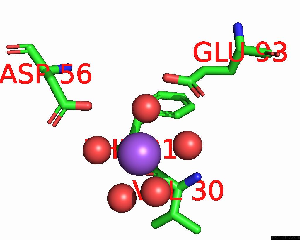
Sodium binding site 1 out of 4 in 7w52
Go back to
Sodium binding site 1 out
of 4 in the Crystal Structure of Fragmin Domain-1 (15-160) in Complex with Actin
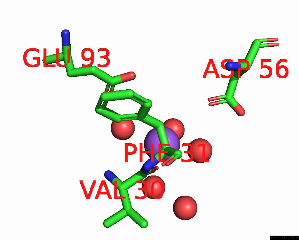
Mono view
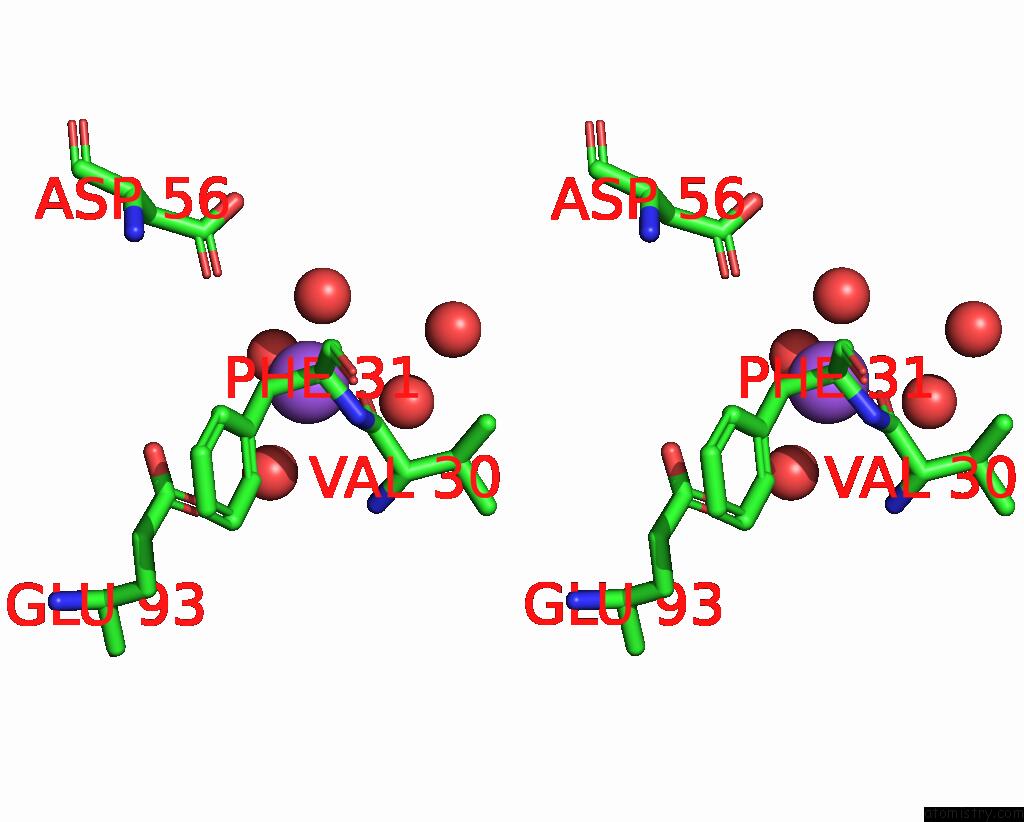
Stereo pair view
Mono view
Stereo pair view
A full contact list of Sodium with other atoms in the Na binding
site number 1 of Crystal Structure of Fragmin Domain-1 (15-160) in Complex with Actin within 5.0Å range:
|
Sodium binding site 2 out of 4 in 7w52
Go back to
Sodium binding site 2 out
of 4 in the Crystal Structure of Fragmin Domain-1 (15-160) in Complex with Actin
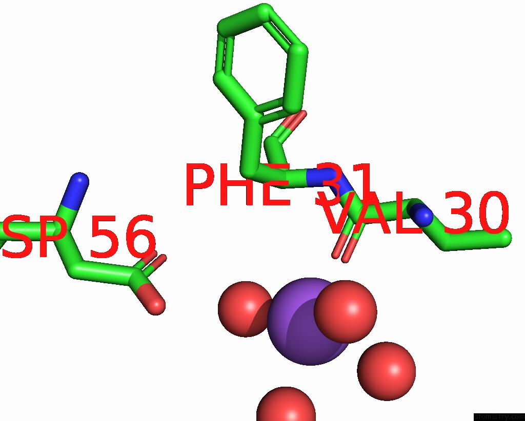
Mono view
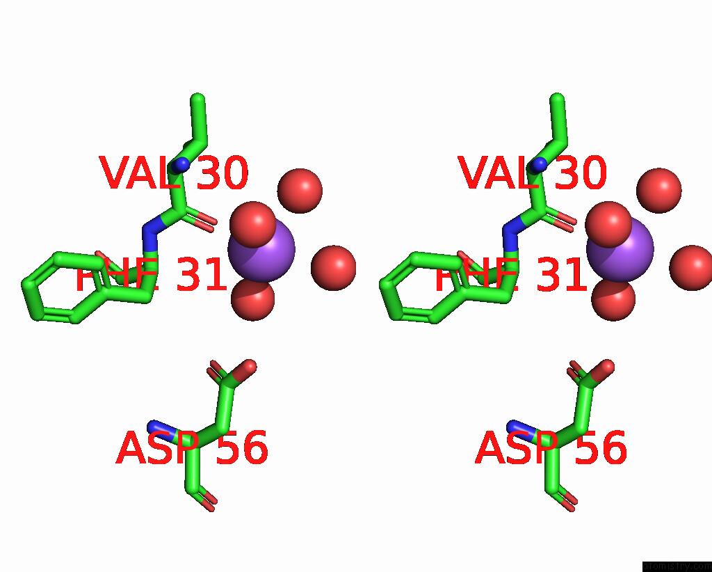
Stereo pair view
Mono view
Stereo pair view
A full contact list of Sodium with other atoms in the Na binding
site number 2 of Crystal Structure of Fragmin Domain-1 (15-160) in Complex with Actin within 5.0Å range:
|
Sodium binding site 3 out of 4 in 7w52
Go back to
Sodium binding site 3 out
of 4 in the Crystal Structure of Fragmin Domain-1 (15-160) in Complex with Actin
Mono view

Stereo pair view
Mono view
Stereo pair view
A full contact list of Sodium with other atoms in the Na binding
site number 3 of Crystal Structure of Fragmin Domain-1 (15-160) in Complex with Actin within 5.0Å range:
|
Sodium binding site 4 out of 4 in 7w52
Go back to
Sodium binding site 4 out
of 4 in the Crystal Structure of Fragmin Domain-1 (15-160) in Complex with Actin

Mono view

Stereo pair view
Mono view
Stereo pair view
A full contact list of Sodium with other atoms in the Na binding
site number 4 of Crystal Structure of Fragmin Domain-1 (15-160) in Complex with Actin within 5.0Å range:
|
Reference:
Y.Kanematsu,
A.Narita,
T.Oda,
R.Koike,
M.Ota,
Y.Takano,
K.Moritsugu,
I.Fujiwara,
K.Tanaka,
H.Komatsu,
T.Nagae,
N.Watanabe,
M.Iwasa,
Y.Maeda,
S.Takeda.
Structures and Mechanisms of Actin Atp Hydrolysis. Proc.Natl.Acad.Sci.Usa V. 119 41119 2022.
ISSN: ESSN 1091-6490
PubMed: 36252034
DOI: 10.1073/PNAS.2122641119
Page generated: Mon Aug 18 12:22:58 2025
ISSN: ESSN 1091-6490
PubMed: 36252034
DOI: 10.1073/PNAS.2122641119
Last articles
Mn in 9LJUMn in 9LJW
Mn in 9LJS
Mn in 9LJR
Mn in 9LJT
Mn in 9LJV
Mg in 9UA2
Mg in 9R96
Mg in 9VM1
Mg in 9P01