Sodium »
PDB 7s79-7smz »
7smz »
Sodium in PDB 7smz: X-Ray Crystal Structure of CYP142A3 From Mycobacterium Marinum in Complex with 4-Cholesten-3-One
Protein crystallography data
The structure of X-Ray Crystal Structure of CYP142A3 From Mycobacterium Marinum in Complex with 4-Cholesten-3-One, PDB code: 7smz
was solved by
A.Ghith,
J.B.Bruning,
S.G.Bell,
with X-Ray Crystallography technique. A brief refinement statistics is given in the table below:
| Resolution Low / High (Å) | 46.69 / 2.04 |
| Space group | P 1 |
| Cell size a, b, c (Å), α, β, γ (°) | 50.313, 50.395, 82.577, 82.32, 75.59, 72.05 |
| R / Rfree (%) | 18 / 22.5 |
Other elements in 7smz:
The structure of X-Ray Crystal Structure of CYP142A3 From Mycobacterium Marinum in Complex with 4-Cholesten-3-One also contains other interesting chemical elements:
| Iron | (Fe) | 2 atoms |
Sodium Binding Sites:
The binding sites of Sodium atom in the X-Ray Crystal Structure of CYP142A3 From Mycobacterium Marinum in Complex with 4-Cholesten-3-One
(pdb code 7smz). This binding sites where shown within
5.0 Angstroms radius around Sodium atom.
In total 4 binding sites of Sodium where determined in the X-Ray Crystal Structure of CYP142A3 From Mycobacterium Marinum in Complex with 4-Cholesten-3-One, PDB code: 7smz:
Jump to Sodium binding site number: 1; 2; 3; 4;
In total 4 binding sites of Sodium where determined in the X-Ray Crystal Structure of CYP142A3 From Mycobacterium Marinum in Complex with 4-Cholesten-3-One, PDB code: 7smz:
Jump to Sodium binding site number: 1; 2; 3; 4;
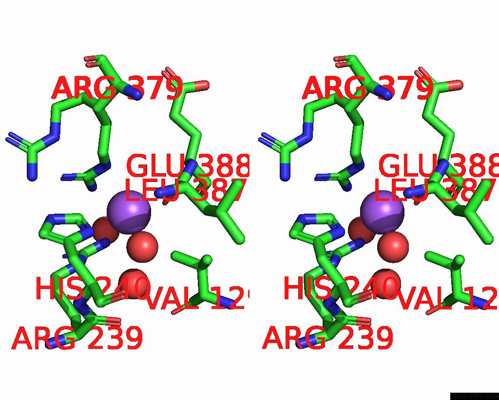
Sodium binding site 1 out of 4 in 7smz
Go back to
Sodium binding site 1 out
of 4 in the X-Ray Crystal Structure of CYP142A3 From Mycobacterium Marinum in Complex with 4-Cholesten-3-One
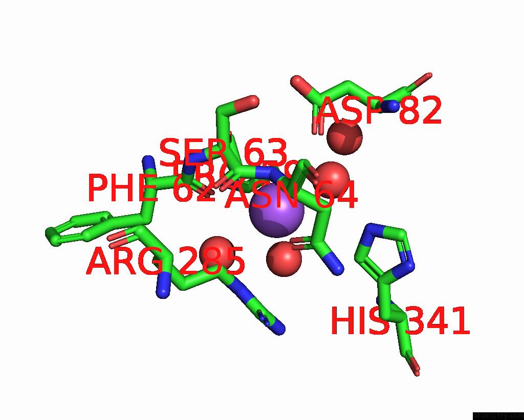
Mono view

Stereo pair view
Mono view
Stereo pair view
A full contact list of Sodium with other atoms in the Na binding
site number 1 of X-Ray Crystal Structure of CYP142A3 From Mycobacterium Marinum in Complex with 4-Cholesten-3-One within 5.0Å range:
|
Sodium binding site 2 out of 4 in 7smz
Go back to
Sodium binding site 2 out
of 4 in the X-Ray Crystal Structure of CYP142A3 From Mycobacterium Marinum in Complex with 4-Cholesten-3-One
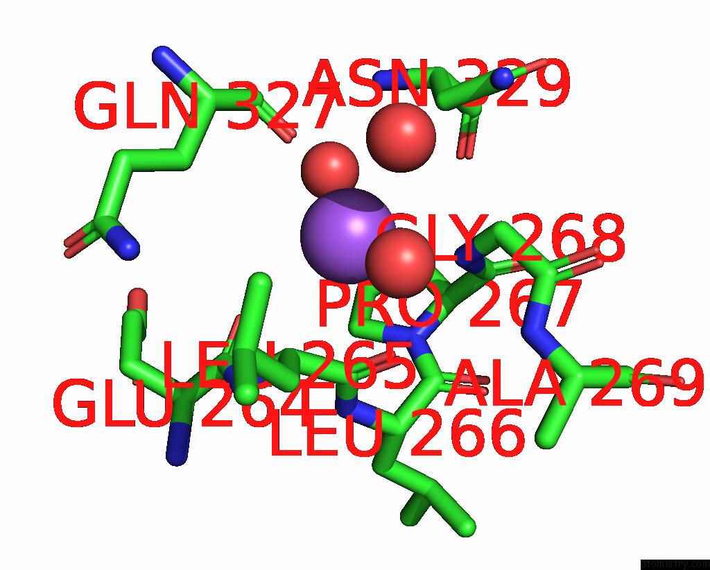
Mono view

Stereo pair view
Mono view
Stereo pair view
A full contact list of Sodium with other atoms in the Na binding
site number 2 of X-Ray Crystal Structure of CYP142A3 From Mycobacterium Marinum in Complex with 4-Cholesten-3-One within 5.0Å range:
|
Sodium binding site 3 out of 4 in 7smz
Go back to
Sodium binding site 3 out
of 4 in the X-Ray Crystal Structure of CYP142A3 From Mycobacterium Marinum in Complex with 4-Cholesten-3-One

Mono view
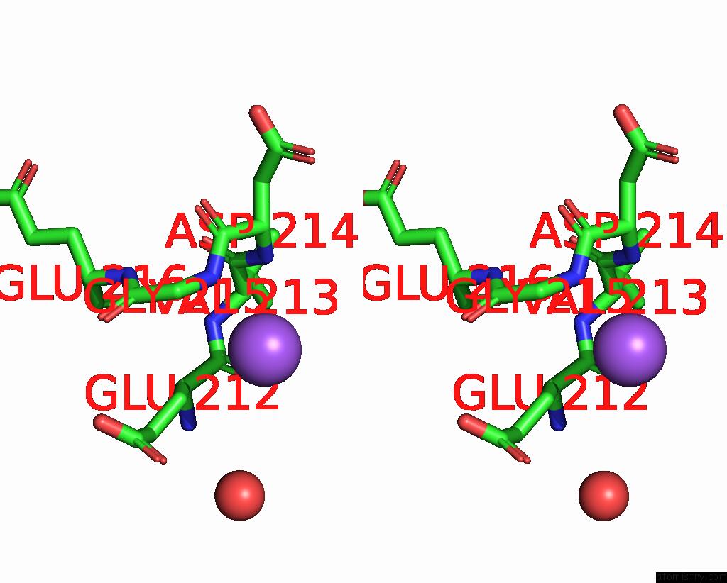
Stereo pair view
Mono view
Stereo pair view
A full contact list of Sodium with other atoms in the Na binding
site number 3 of X-Ray Crystal Structure of CYP142A3 From Mycobacterium Marinum in Complex with 4-Cholesten-3-One within 5.0Å range:
|
Sodium binding site 4 out of 4 in 7smz
Go back to
Sodium binding site 4 out
of 4 in the X-Ray Crystal Structure of CYP142A3 From Mycobacterium Marinum in Complex with 4-Cholesten-3-One
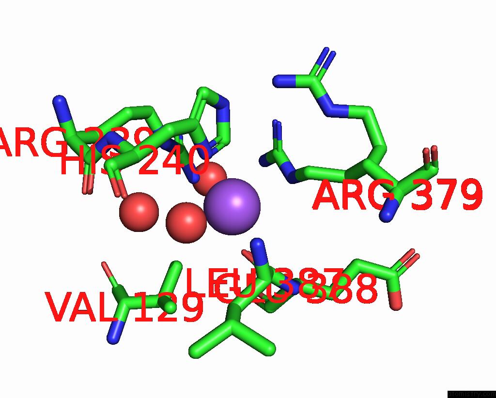
Mono view
Stereo pair view
Mono view
Stereo pair view
A full contact list of Sodium with other atoms in the Na binding
site number 4 of X-Ray Crystal Structure of CYP142A3 From Mycobacterium Marinum in Complex with 4-Cholesten-3-One within 5.0Å range:
|
Reference:
A.Ghith,
D.Z.Doherty,
J.B.Bruning,
R.A.Russell,
J.J.De Voss,
S.G.Bell.
The Structures of the Steroid Binding CYP142 Cytochrome P450 Enzymes From Mycobacterium Ulcerans and Mycobacterium Marinum. Acs Infect Dis. V. 8 1606 2022.
ISSN: ESSN 2373-8227
PubMed: 35881654
DOI: 10.1021/ACSINFECDIS.2C00215
Page generated: Mon Aug 18 11:58:52 2025
ISSN: ESSN 2373-8227
PubMed: 35881654
DOI: 10.1021/ACSINFECDIS.2C00215
Last articles
Mn in 9LJUMn in 9LJW
Mn in 9LJS
Mn in 9LJR
Mn in 9LJT
Mn in 9LJV
Mg in 9UA2
Mg in 9R96
Mg in 9VM1
Mg in 9P01