Sodium »
PDB 7bqj-7cdu »
7c9b »
Sodium in PDB 7c9b: Crystal Structure of Di-Peptidase-E From Xenopus Laevis
Enzymatic activity of Crystal Structure of Di-Peptidase-E From Xenopus Laevis
All present enzymatic activity of Crystal Structure of Di-Peptidase-E From Xenopus Laevis:
3.4.13.21;
3.4.13.21;
Protein crystallography data
The structure of Crystal Structure of Di-Peptidase-E From Xenopus Laevis, PDB code: 7c9b
was solved by
A.Kumar,
R.Singh,
R.D.Makde,
with X-Ray Crystallography technique. A brief refinement statistics is given in the table below:
| Resolution Low / High (Å) | 34.84 / 1.40 |
| Space group | C 1 2 1 |
| Cell size a, b, c (Å), α, β, γ (°) | 83.775, 40.9, 74.221, 90, 116.2, 90 |
| R / Rfree (%) | 20 / 22.6 |
Other elements in 7c9b:
The structure of Crystal Structure of Di-Peptidase-E From Xenopus Laevis also contains other interesting chemical elements:
| Calcium | (Ca) | 1 atom |
Sodium Binding Sites:
The binding sites of Sodium atom in the Crystal Structure of Di-Peptidase-E From Xenopus Laevis
(pdb code 7c9b). This binding sites where shown within
5.0 Angstroms radius around Sodium atom.
In total only one binding site of Sodium was determined in the Crystal Structure of Di-Peptidase-E From Xenopus Laevis, PDB code: 7c9b:
In total only one binding site of Sodium was determined in the Crystal Structure of Di-Peptidase-E From Xenopus Laevis, PDB code: 7c9b:
Sodium binding site 1 out of 1 in 7c9b
Go back to
Sodium binding site 1 out
of 1 in the Crystal Structure of Di-Peptidase-E From Xenopus Laevis

Mono view
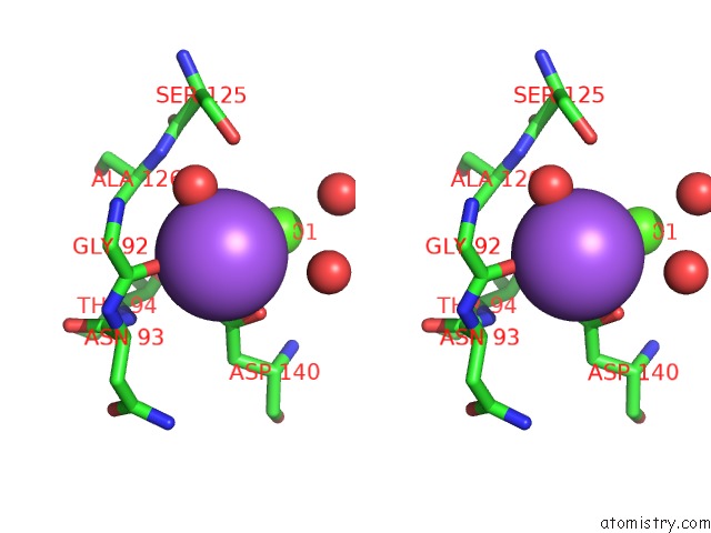
Stereo pair view
Mono view
Stereo pair view
A full contact list of Sodium with other atoms in the Na binding
site number 1 of Crystal Structure of Di-Peptidase-E From Xenopus Laevis within 5.0Å range:
|
Reference:
A.Kumar,
R.Singh,
R.D.Makde.
Crystal Structure of Di-Peptidase-E From Xenopus Laevis To Be Published.
Page generated: Tue Oct 8 16:19:59 2024
Last articles
Mg in 1KXPMg in 1KXK
Mg in 1KTG
Mg in 1KWO
Mg in 1KW1
Mg in 1KP8
Mg in 1KVK
Mg in 1KV8
Mg in 1KUV
Mg in 1KSZ