Sodium »
PDB 6vf7-6w0r »
6vut »
Sodium in PDB 6vut: Crystal Structure of Eis From Mycobacterium Tuberculosis in Complex with Inhibitor SGT392
Protein crystallography data
The structure of Crystal Structure of Eis From Mycobacterium Tuberculosis in Complex with Inhibitor SGT392, PDB code: 6vut
was solved by
A.Punetha,
C.Hou,
H.X.Ngo,
S.Garneau-Tsodikova,
O.V.Tsodikov,
with X-Ray Crystallography technique. A brief refinement statistics is given in the table below:
| Resolution Low / High (Å) | 36.96 / 2.73 |
| Space group | H 3 2 |
| Cell size a, b, c (Å), α, β, γ (°) | 175.190, 175.190, 122.191, 90.00, 90.00, 120.00 |
| R / Rfree (%) | 18.3 / 22.7 |
Sodium Binding Sites:
The binding sites of Sodium atom in the Crystal Structure of Eis From Mycobacterium Tuberculosis in Complex with Inhibitor SGT392
(pdb code 6vut). This binding sites where shown within
5.0 Angstroms radius around Sodium atom.
In total 3 binding sites of Sodium where determined in the Crystal Structure of Eis From Mycobacterium Tuberculosis in Complex with Inhibitor SGT392, PDB code: 6vut:
Jump to Sodium binding site number: 1; 2; 3;
In total 3 binding sites of Sodium where determined in the Crystal Structure of Eis From Mycobacterium Tuberculosis in Complex with Inhibitor SGT392, PDB code: 6vut:
Jump to Sodium binding site number: 1; 2; 3;
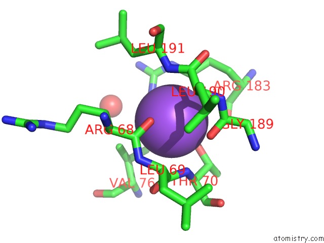
Sodium binding site 1 out of 3 in 6vut
Go back to
Sodium binding site 1 out
of 3 in the Crystal Structure of Eis From Mycobacterium Tuberculosis in Complex with Inhibitor SGT392
Mono view

Stereo pair view
Mono view
Stereo pair view
A full contact list of Sodium with other atoms in the Na binding
site number 1 of Crystal Structure of Eis From Mycobacterium Tuberculosis in Complex with Inhibitor SGT392 within 5.0Å range:
|
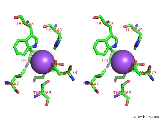
Sodium binding site 2 out of 3 in 6vut
Go back to
Sodium binding site 2 out
of 3 in the Crystal Structure of Eis From Mycobacterium Tuberculosis in Complex with Inhibitor SGT392

Mono view
Stereo pair view
Mono view
Stereo pair view
A full contact list of Sodium with other atoms in the Na binding
site number 2 of Crystal Structure of Eis From Mycobacterium Tuberculosis in Complex with Inhibitor SGT392 within 5.0Å range:
|
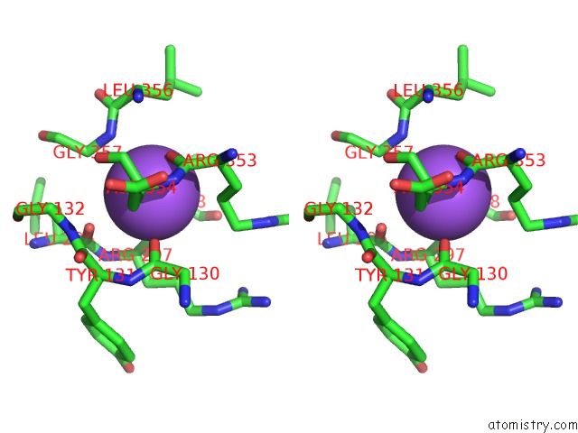
Sodium binding site 3 out of 3 in 6vut
Go back to
Sodium binding site 3 out
of 3 in the Crystal Structure of Eis From Mycobacterium Tuberculosis in Complex with Inhibitor SGT392

Mono view
Stereo pair view
Mono view
Stereo pair view
A full contact list of Sodium with other atoms in the Na binding
site number 3 of Crystal Structure of Eis From Mycobacterium Tuberculosis in Complex with Inhibitor SGT392 within 5.0Å range:
|
Reference:
A.Punetha,
H.X.Ngo,
S.Y.L.Holbrook,
K.D.Green,
M.J.Willby,
S.A.Bonnett,
K.Krieger,
E.K.Dennis,
J.E.Posey,
T.Parish,
O.V.Tsodikov,
S.Garneau-Tsodikova.
Structure-Guided Optimization of Inhibitors of Acetyltransferase Eis Frommycobacterium Tuberculosis. Acs Chem.Biol. 2020.
ISSN: ESSN 1554-8937
PubMed: 32421305
DOI: 10.1021/ACSCHEMBIO.0C00184
Page generated: Tue Oct 8 14:26:22 2024
ISSN: ESSN 1554-8937
PubMed: 32421305
DOI: 10.1021/ACSCHEMBIO.0C00184
Last articles
Mg in 6L5NMg in 6L59
Mg in 6L5L
Mg in 6L4N
Mg in 6L57
Mg in 6L54
Mg in 6L3R
Mg in 6L42
Mg in 6L3E
Mg in 6L3G