Sodium »
PDB 6tbt-6tos »
6tos »
Sodium in PDB 6tos: Crystal Structure of the Orexin-1 Receptor in Complex with GSK1059865
Protein crystallography data
The structure of Crystal Structure of the Orexin-1 Receptor in Complex with GSK1059865, PDB code: 6tos
was solved by
M.Rappas,
A.Ali,
K.A.Bennett,
J.D.Brown,
S.J.Bucknell,
M.Congreve,
R.M.Cooke,
G.Cseke,
C.De Graaf,
A.S.Dore,
J.C.Errey,
A.Jazayeri,
F.H.Marshall,
J.S.Mason,
R.Mould,
J.C.Patel,
B.G.Tehan,
M.Weir,
J.A.Christopher,
with X-Ray Crystallography technique. A brief refinement statistics is given in the table below:
| Resolution Low / High (Å) | 30.47 / 2.13 |
| Space group | P 1 21 1 |
| Cell size a, b, c (Å), α, β, γ (°) | 59.692, 146.190, 71.508, 90.00, 112.43, 90.00 |
| R / Rfree (%) | 19.3 / 21.8 |
Other elements in 6tos:
The structure of Crystal Structure of the Orexin-1 Receptor in Complex with GSK1059865 also contains other interesting chemical elements:
| Fluorine | (F) | 2 atoms |
| Bromine | (Br) | 2 atoms |
Sodium Binding Sites:
The binding sites of Sodium atom in the Crystal Structure of the Orexin-1 Receptor in Complex with GSK1059865
(pdb code 6tos). This binding sites where shown within
5.0 Angstroms radius around Sodium atom.
In total only one binding site of Sodium was determined in the Crystal Structure of the Orexin-1 Receptor in Complex with GSK1059865, PDB code: 6tos:
In total only one binding site of Sodium was determined in the Crystal Structure of the Orexin-1 Receptor in Complex with GSK1059865, PDB code: 6tos:
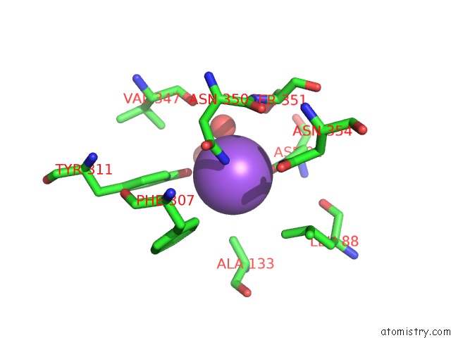
Sodium binding site 1 out of 1 in 6tos
Go back to
Sodium binding site 1 out
of 1 in the Crystal Structure of the Orexin-1 Receptor in Complex with GSK1059865
Mono view

Stereo pair view
Mono view
Stereo pair view
A full contact list of Sodium with other atoms in the Na binding
site number 1 of Crystal Structure of the Orexin-1 Receptor in Complex with GSK1059865 within 5.0Å range:
|
Reference:
M.Rappas,
A.Ali,
K.A.Bennett,
J.D.Brown,
S.J.Bucknell,
M.Congreve,
R.M.Cooke,
G.Cseke,
C.De Graaf,
A.S.Dore,
J.C.Errey,
A.Jazayeri,
F.H.Marshall,
J.S.Mason,
R.Mould,
J.C.Patel,
B.Tehan,
M.Weir,
J.A.Christopher.
Comparison of Orexin 1 and Orexin 2 Ligand Binding Modes Using X-Ray Crystallography and Computational Analysis. J.Med.Chem. 2019.
ISSN: ISSN 0022-2623
PubMed: 31860301
DOI: 10.1021/ACS.JMEDCHEM.9B01787
Page generated: Tue Oct 8 14:00:12 2024
ISSN: ISSN 0022-2623
PubMed: 31860301
DOI: 10.1021/ACS.JMEDCHEM.9B01787
Last articles
Mg in 4QWLMg in 4QWR
Mg in 4QWK
Mg in 4QWJ
Mg in 4QWI
Mg in 4QWF
Mg in 4QWG
Mg in 4QW7
Mg in 4QW6
Mg in 4QW4