Sodium »
PDB 6kcd-6lgy »
6kgh »
Sodium in PDB 6kgh: Crystal Structure of Penicillin Binding Protein 3 (PBP3) From Mycobacterium Tuerculosis (Apo-Form)
Protein crystallography data
The structure of Crystal Structure of Penicillin Binding Protein 3 (PBP3) From Mycobacterium Tuerculosis (Apo-Form), PDB code: 6kgh
was solved by
Z.K.Lu,
A.L.Zhang,
X.Liu,
L.Guddat,
H.T.Yang,
Z.H.Rao,
with X-Ray Crystallography technique. A brief refinement statistics is given in the table below:
| Resolution Low / High (Å) | 48.04 / 2.11 |
| Space group | C 1 2 1 |
| Cell size a, b, c (Å), α, β, γ (°) | 97.710, 84.220, 90.500, 90.00, 111.60, 90.00 |
| R / Rfree (%) | 18.9 / 22.2 |
Other elements in 6kgh:
The structure of Crystal Structure of Penicillin Binding Protein 3 (PBP3) From Mycobacterium Tuerculosis (Apo-Form) also contains other interesting chemical elements:
| Cobalt | (Co) | 3 atoms |
Sodium Binding Sites:
The binding sites of Sodium atom in the Crystal Structure of Penicillin Binding Protein 3 (PBP3) From Mycobacterium Tuerculosis (Apo-Form)
(pdb code 6kgh). This binding sites where shown within
5.0 Angstroms radius around Sodium atom.
In total only one binding site of Sodium was determined in the Crystal Structure of Penicillin Binding Protein 3 (PBP3) From Mycobacterium Tuerculosis (Apo-Form), PDB code: 6kgh:
In total only one binding site of Sodium was determined in the Crystal Structure of Penicillin Binding Protein 3 (PBP3) From Mycobacterium Tuerculosis (Apo-Form), PDB code: 6kgh:
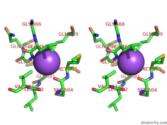
Sodium binding site 1 out of 1 in 6kgh
Go back to
Sodium binding site 1 out
of 1 in the Crystal Structure of Penicillin Binding Protein 3 (PBP3) From Mycobacterium Tuerculosis (Apo-Form)

Mono view
Stereo pair view
Mono view
Stereo pair view
A full contact list of Sodium with other atoms in the Na binding
site number 1 of Crystal Structure of Penicillin Binding Protein 3 (PBP3) From Mycobacterium Tuerculosis (Apo-Form) within 5.0Å range:
|
Reference:
Z.Lu,
H.Wang,
A.Zhang,
X.Liu,
W.Zhou,
C.Yang,
L.Guddat,
H.Yang,
C.J.Schofield,
Z.Rao.
Structures Ofmycobacterium Tuberculosispenicillin-Binding Protein 3 in Complex with Fivebeta-Lactam Antibiotics Reveal Mechanism of Inactivation. Mol.Pharmacol. V. 97 287 2020.
ISSN: ESSN 1521-0111
PubMed: 32086254
DOI: 10.1124/MOL.119.118042
Page generated: Mon Aug 18 05:41:13 2025
ISSN: ESSN 1521-0111
PubMed: 32086254
DOI: 10.1124/MOL.119.118042
Last articles
Na in 7G4MNa in 7G4L
Na in 7G4H
Na in 7G4K
Na in 7G49
Na in 7G4B
Na in 7G48
Na in 7G47
Na in 7G46
Na in 7G45