Sodium »
PDB 6hja-6hz0 »
6hk1 »
Sodium in PDB 6hk1: Crystal Structure of the Thiazole Synthase From Methanothermococcus Thermolithotrophicus Co-Crystallized with Tb-XO4
Protein crystallography data
The structure of Crystal Structure of the Thiazole Synthase From Methanothermococcus Thermolithotrophicus Co-Crystallized with Tb-XO4, PDB code: 6hk1
was solved by
S.Engilberge,
T.Wagner,
G.Santoni,
C.Breyton,
S.Shima,
B.Franzetti,
F.Riobe,
O.Maury,
E.Girard,
with X-Ray Crystallography technique. A brief refinement statistics is given in the table below:
| Resolution Low / High (Å) | 49.09 / 2.55 |
| Space group | I 4 2 2 |
| Cell size a, b, c (Å), α, β, γ (°) | 216.863, 216.863, 207.250, 90.00, 90.00, 90.00 |
| R / Rfree (%) | 18.9 / 21.3 |
Other elements in 6hk1:
The structure of Crystal Structure of the Thiazole Synthase From Methanothermococcus Thermolithotrophicus Co-Crystallized with Tb-XO4 also contains other interesting chemical elements:
| Terbium | (Tb) | 49 atoms |
Sodium Binding Sites:
The binding sites of Sodium atom in the Crystal Structure of the Thiazole Synthase From Methanothermococcus Thermolithotrophicus Co-Crystallized with Tb-XO4
(pdb code 6hk1). This binding sites where shown within
5.0 Angstroms radius around Sodium atom.
In total only one binding site of Sodium was determined in the Crystal Structure of the Thiazole Synthase From Methanothermococcus Thermolithotrophicus Co-Crystallized with Tb-XO4, PDB code: 6hk1:
In total only one binding site of Sodium was determined in the Crystal Structure of the Thiazole Synthase From Methanothermococcus Thermolithotrophicus Co-Crystallized with Tb-XO4, PDB code: 6hk1:
Sodium binding site 1 out of 1 in 6hk1
Go back to
Sodium binding site 1 out
of 1 in the Crystal Structure of the Thiazole Synthase From Methanothermococcus Thermolithotrophicus Co-Crystallized with Tb-XO4
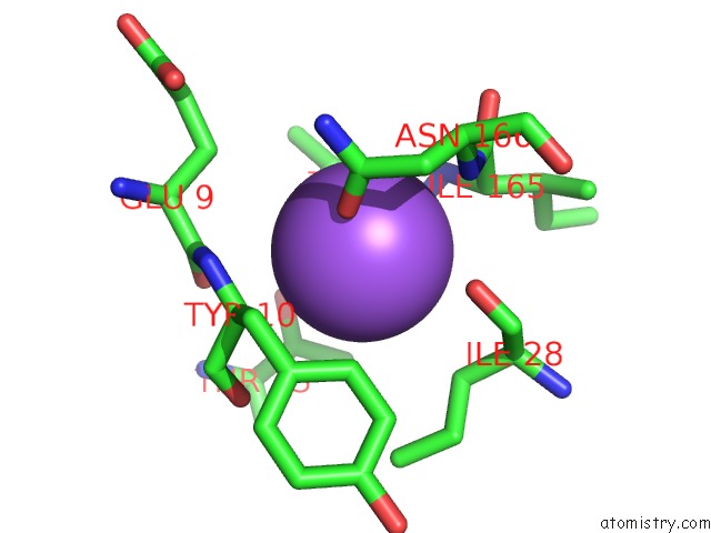
Mono view
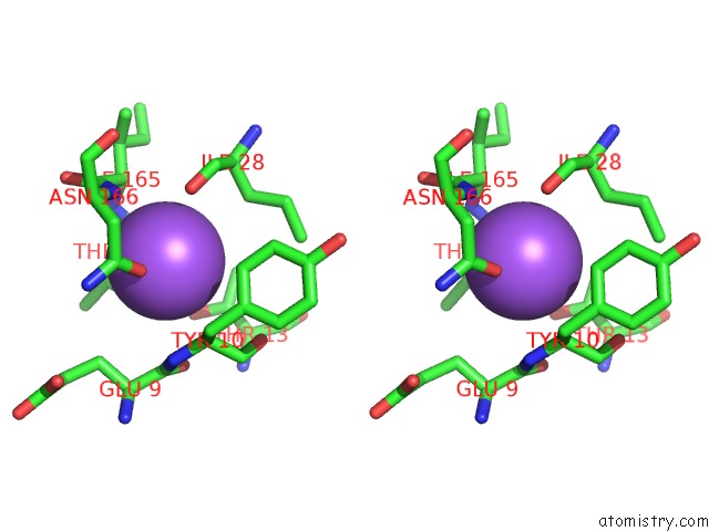
Stereo pair view
Mono view
Stereo pair view
A full contact list of Sodium with other atoms in the Na binding
site number 1 of Crystal Structure of the Thiazole Synthase From Methanothermococcus Thermolithotrophicus Co-Crystallized with Tb-XO4 within 5.0Å range:
|
Reference:
S.Engilberge,
T.Wagner,
G.Santoni,
C.Breyton,
S.Shima,
B.Franzetti,
F.Riobe,
O.Maury,
E.Girard.
Protein Crystal Structure Determination with the Crystallophore, A Nucleating and Phasing Agent. J.Appl.Crystallogr. V. 52 722 2019.
ISSN: ISSN 0021-8898
PubMed: 31396026
DOI: 10.1107/S1600576719006381
Page generated: Mon Aug 18 05:16:58 2025
ISSN: ISSN 0021-8898
PubMed: 31396026
DOI: 10.1107/S1600576719006381
Last articles
Mn in 9LJUMn in 9LJW
Mn in 9LJS
Mn in 9LJR
Mn in 9LJT
Mn in 9LJV
Mg in 9UA2
Mg in 9R96
Mg in 9VM1
Mg in 9P01