Sodium »
PDB 5xvd-5ysu »
5ydb »
Sodium in PDB 5ydb: Crystal Structure of the Complex of Type II Dehydroquinate Dehydratase From Acinetobacter Baumannii with Dehydroquinic Acid at 1.76 Angstrom Resolution
Enzymatic activity of Crystal Structure of the Complex of Type II Dehydroquinate Dehydratase From Acinetobacter Baumannii with Dehydroquinic Acid at 1.76 Angstrom Resolution
All present enzymatic activity of Crystal Structure of the Complex of Type II Dehydroquinate Dehydratase From Acinetobacter Baumannii with Dehydroquinic Acid at 1.76 Angstrom Resolution:
4.2.1.10;
4.2.1.10;
Protein crystallography data
The structure of Crystal Structure of the Complex of Type II Dehydroquinate Dehydratase From Acinetobacter Baumannii with Dehydroquinic Acid at 1.76 Angstrom Resolution, PDB code: 5ydb
was solved by
N.Iqbal,
P.Kaur,
S.Sharma,
T.P.Singh,
with X-Ray Crystallography technique. A brief refinement statistics is given in the table below:
| Resolution Low / High (Å) | 69.56 / 1.76 |
| Space group | H 3 |
| Cell size a, b, c (Å), α, β, γ (°) | 139.117, 139.117, 79.587, 90.00, 90.00, 120.00 |
| R / Rfree (%) | 19.7 / 24.8 |
Sodium Binding Sites:
The binding sites of Sodium atom in the Crystal Structure of the Complex of Type II Dehydroquinate Dehydratase From Acinetobacter Baumannii with Dehydroquinic Acid at 1.76 Angstrom Resolution
(pdb code 5ydb). This binding sites where shown within
5.0 Angstroms radius around Sodium atom.
In total 4 binding sites of Sodium where determined in the Crystal Structure of the Complex of Type II Dehydroquinate Dehydratase From Acinetobacter Baumannii with Dehydroquinic Acid at 1.76 Angstrom Resolution, PDB code: 5ydb:
Jump to Sodium binding site number: 1; 2; 3; 4;
In total 4 binding sites of Sodium where determined in the Crystal Structure of the Complex of Type II Dehydroquinate Dehydratase From Acinetobacter Baumannii with Dehydroquinic Acid at 1.76 Angstrom Resolution, PDB code: 5ydb:
Jump to Sodium binding site number: 1; 2; 3; 4;
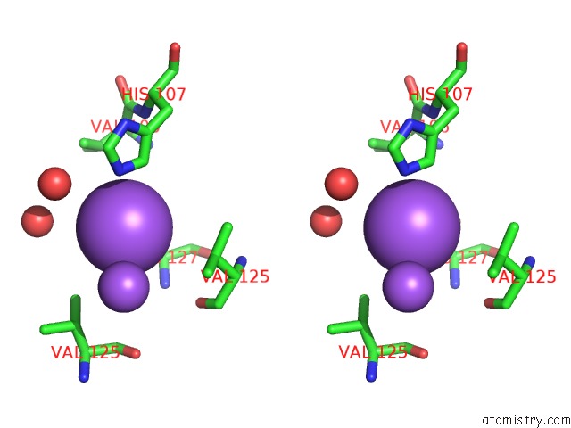
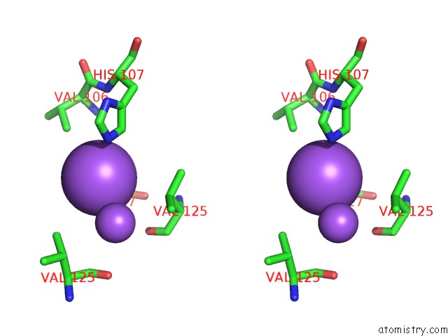
Sodium binding site 1 out of 4 in 5ydb
Go back to
Sodium binding site 1 out
of 4 in the Crystal Structure of the Complex of Type II Dehydroquinate Dehydratase From Acinetobacter Baumannii with Dehydroquinic Acid at 1.76 Angstrom Resolution
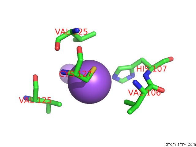
Mono view

Stereo pair view
Mono view
Stereo pair view
A full contact list of Sodium with other atoms in the Na binding
site number 1 of Crystal Structure of the Complex of Type II Dehydroquinate Dehydratase From Acinetobacter Baumannii with Dehydroquinic Acid at 1.76 Angstrom Resolution within 5.0Å range:
|
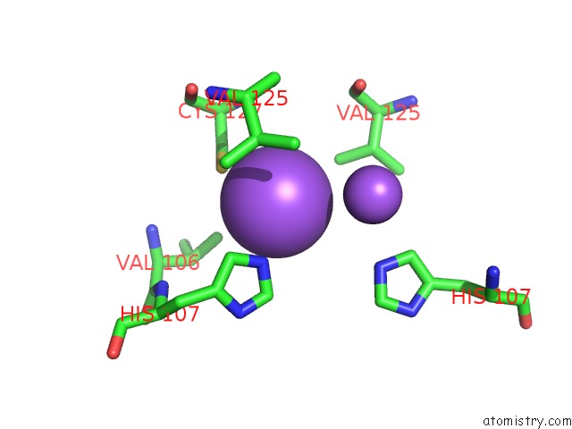
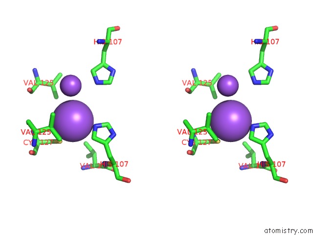
Sodium binding site 2 out of 4 in 5ydb
Go back to
Sodium binding site 2 out
of 4 in the Crystal Structure of the Complex of Type II Dehydroquinate Dehydratase From Acinetobacter Baumannii with Dehydroquinic Acid at 1.76 Angstrom Resolution

Mono view
Stereo pair view
Mono view
Stereo pair view
A full contact list of Sodium with other atoms in the Na binding
site number 2 of Crystal Structure of the Complex of Type II Dehydroquinate Dehydratase From Acinetobacter Baumannii with Dehydroquinic Acid at 1.76 Angstrom Resolution within 5.0Å range:
|
Sodium binding site 3 out of 4 in 5ydb
Go back to
Sodium binding site 3 out
of 4 in the Crystal Structure of the Complex of Type II Dehydroquinate Dehydratase From Acinetobacter Baumannii with Dehydroquinic Acid at 1.76 Angstrom Resolution
Mono view
Stereo pair view
Mono view
Stereo pair view
A full contact list of Sodium with other atoms in the Na binding
site number 3 of Crystal Structure of the Complex of Type II Dehydroquinate Dehydratase From Acinetobacter Baumannii with Dehydroquinic Acid at 1.76 Angstrom Resolution within 5.0Å range:
|
Sodium binding site 4 out of 4 in 5ydb
Go back to
Sodium binding site 4 out
of 4 in the Crystal Structure of the Complex of Type II Dehydroquinate Dehydratase From Acinetobacter Baumannii with Dehydroquinic Acid at 1.76 Angstrom Resolution

Mono view
Stereo pair view
Mono view
Stereo pair view
A full contact list of Sodium with other atoms in the Na binding
site number 4 of Crystal Structure of the Complex of Type II Dehydroquinate Dehydratase From Acinetobacter Baumannii with Dehydroquinic Acid at 1.76 Angstrom Resolution within 5.0Å range:
|
Reference:
N.Iqbal,
P.Kaur,
S.Sharma,
T.P.Singh.
Crystal Structure of the Complex of Type II Dehydroquinate Dehydratase From Acinetobacter Baumannii with Dehydroquinic Acid at 1.76 Angstrom Resolution To Be Published.
Page generated: Tue Oct 8 01:22:23 2024
Last articles
Fe in 6M0AFe in 6LVV
Fe in 6LU1
Fe in 6LY4
Fe in 6LVC
Fe in 6LVB
Fe in 6LS3
Fe in 6LS8
Fe in 6LTM
Fe in 6LTL