Sodium »
PDB 5vbx-5vxa »
5vev »
Sodium in PDB 5vev: Crystal Structure of Phosphoribosylamine-Glycine Ligase From Neisseria Gonorrhoeae
Enzymatic activity of Crystal Structure of Phosphoribosylamine-Glycine Ligase From Neisseria Gonorrhoeae
All present enzymatic activity of Crystal Structure of Phosphoribosylamine-Glycine Ligase From Neisseria Gonorrhoeae:
6.3.4.13;
6.3.4.13;
Protein crystallography data
The structure of Crystal Structure of Phosphoribosylamine-Glycine Ligase From Neisseria Gonorrhoeae, PDB code: 5vev
was solved by
Seattle Structural Genomics Center For Infectious Disease (Ssgcid),
with X-Ray Crystallography technique. A brief refinement statistics is given in the table below:
| Resolution Low / High (Å) | 46.52 / 1.90 |
| Space group | P 21 21 21 |
| Cell size a, b, c (Å), α, β, γ (°) | 73.310, 99.450, 131.600, 90.00, 90.00, 90.00 |
| R / Rfree (%) | 16.6 / 20.1 |
Sodium Binding Sites:
The binding sites of Sodium atom in the Crystal Structure of Phosphoribosylamine-Glycine Ligase From Neisseria Gonorrhoeae
(pdb code 5vev). This binding sites where shown within
5.0 Angstroms radius around Sodium atom.
In total 2 binding sites of Sodium where determined in the Crystal Structure of Phosphoribosylamine-Glycine Ligase From Neisseria Gonorrhoeae, PDB code: 5vev:
Jump to Sodium binding site number: 1; 2;
In total 2 binding sites of Sodium where determined in the Crystal Structure of Phosphoribosylamine-Glycine Ligase From Neisseria Gonorrhoeae, PDB code: 5vev:
Jump to Sodium binding site number: 1; 2;
Sodium binding site 1 out of 2 in 5vev
Go back to
Sodium binding site 1 out
of 2 in the Crystal Structure of Phosphoribosylamine-Glycine Ligase From Neisseria Gonorrhoeae
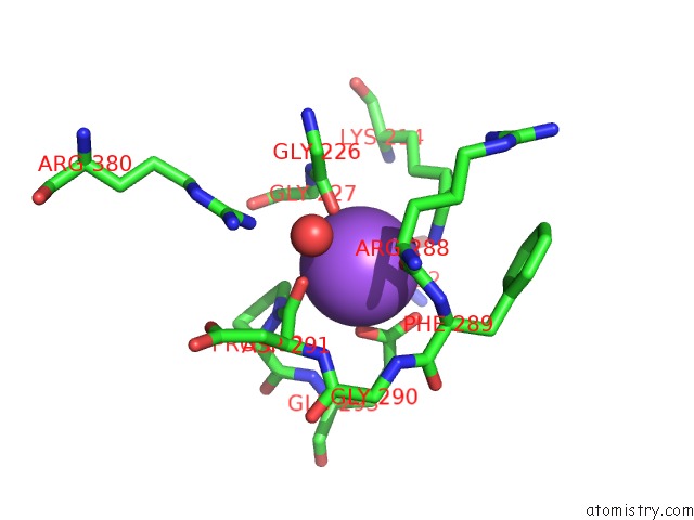
Mono view
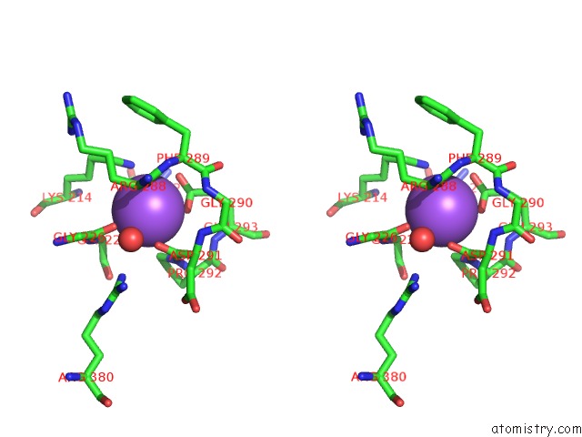
Stereo pair view
Mono view
Stereo pair view
A full contact list of Sodium with other atoms in the Na binding
site number 1 of Crystal Structure of Phosphoribosylamine-Glycine Ligase From Neisseria Gonorrhoeae within 5.0Å range:
|
Sodium binding site 2 out of 2 in 5vev
Go back to
Sodium binding site 2 out
of 2 in the Crystal Structure of Phosphoribosylamine-Glycine Ligase From Neisseria Gonorrhoeae
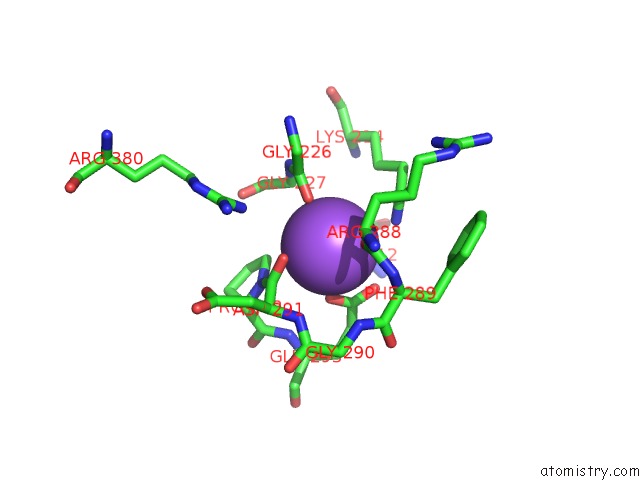
Mono view
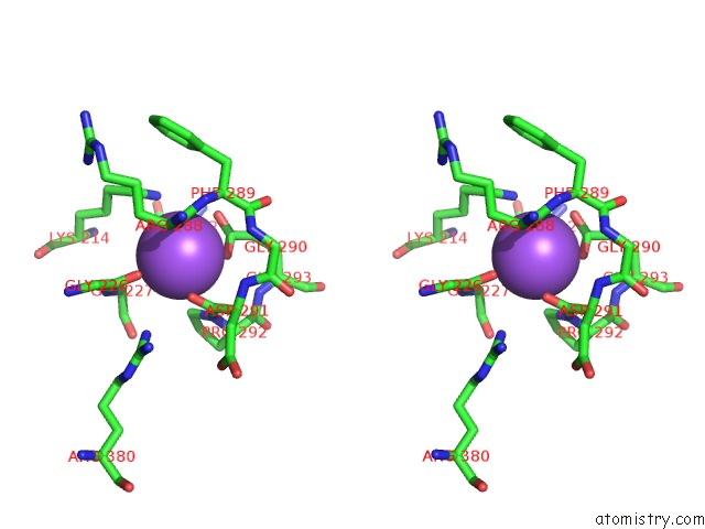
Stereo pair view
Mono view
Stereo pair view
A full contact list of Sodium with other atoms in the Na binding
site number 2 of Crystal Structure of Phosphoribosylamine-Glycine Ligase From Neisseria Gonorrhoeae within 5.0Å range:
|
Reference:
D.M.Dranow,
J.Abendroth,
D.D.Lorimer,
T.E.Edwards.
Crystal Structure of Phosphoribosylamine-Glycine Ligase From Neisseria Gonorrhoeae To Be Published.
Page generated: Tue Oct 8 00:48:02 2024
Last articles
Mg in 3CC7Mg in 3CC4
Mg in 3CB3
Mg in 3CC6
Mg in 3C9U
Mg in 3CBT
Mg in 3CBQ
Mg in 3CBG
Mg in 3CBE
Mg in 3C9T