Sodium »
PDB 5uka-5v0d »
5ur0 »
Sodium in PDB 5ur0: Crystallographic Structure of Glyceraldehyde-3-Phosphate Dehydrogenase From Naegleria Gruberi
Enzymatic activity of Crystallographic Structure of Glyceraldehyde-3-Phosphate Dehydrogenase From Naegleria Gruberi
All present enzymatic activity of Crystallographic Structure of Glyceraldehyde-3-Phosphate Dehydrogenase From Naegleria Gruberi:
1.2.1.12;
1.2.1.12;
Protein crystallography data
The structure of Crystallographic Structure of Glyceraldehyde-3-Phosphate Dehydrogenase From Naegleria Gruberi, PDB code: 5ur0
was solved by
A.T.P.Machado,
M.Silva,
J.Iulek,
with X-Ray Crystallography technique. A brief refinement statistics is given in the table below:
| Resolution Low / High (Å) | 29.85 / 1.94 |
| Space group | P 1 21 1 |
| Cell size a, b, c (Å), α, β, γ (°) | 83.743, 94.552, 90.932, 90.00, 99.96, 90.00 |
| R / Rfree (%) | 15.4 / 19.8 |
Sodium Binding Sites:
The binding sites of Sodium atom in the Crystallographic Structure of Glyceraldehyde-3-Phosphate Dehydrogenase From Naegleria Gruberi
(pdb code 5ur0). This binding sites where shown within
5.0 Angstroms radius around Sodium atom.
In total 4 binding sites of Sodium where determined in the Crystallographic Structure of Glyceraldehyde-3-Phosphate Dehydrogenase From Naegleria Gruberi, PDB code: 5ur0:
Jump to Sodium binding site number: 1; 2; 3; 4;
In total 4 binding sites of Sodium where determined in the Crystallographic Structure of Glyceraldehyde-3-Phosphate Dehydrogenase From Naegleria Gruberi, PDB code: 5ur0:
Jump to Sodium binding site number: 1; 2; 3; 4;
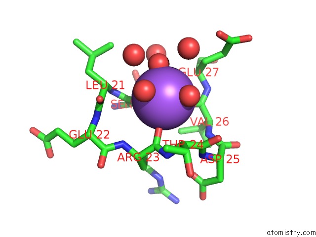
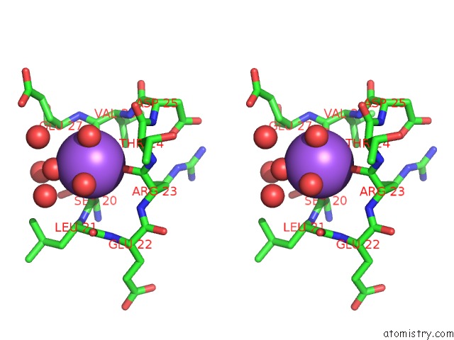
Sodium binding site 1 out of 4 in 5ur0
Go back to
Sodium binding site 1 out
of 4 in the Crystallographic Structure of Glyceraldehyde-3-Phosphate Dehydrogenase From Naegleria Gruberi
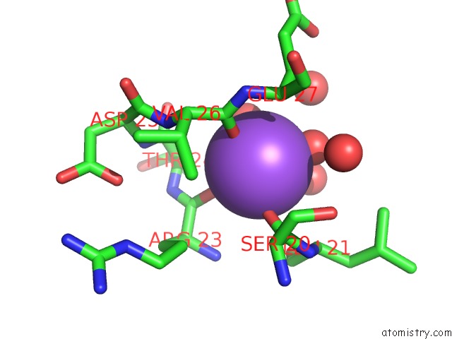
Mono view
Stereo pair view
Mono view
Stereo pair view
A full contact list of Sodium with other atoms in the Na binding
site number 1 of Crystallographic Structure of Glyceraldehyde-3-Phosphate Dehydrogenase From Naegleria Gruberi within 5.0Å range:
|
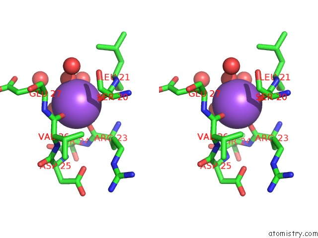
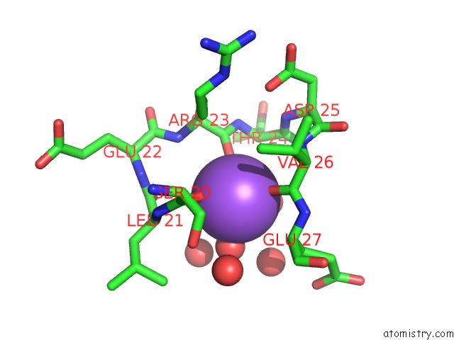
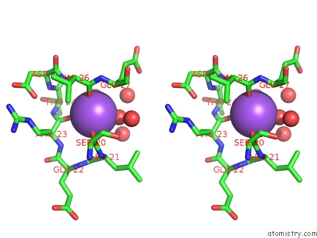
Sodium binding site 2 out of 4 in 5ur0
Go back to
Sodium binding site 2 out
of 4 in the Crystallographic Structure of Glyceraldehyde-3-Phosphate Dehydrogenase From Naegleria Gruberi
Mono view
Stereo pair view
Mono view
Stereo pair view
A full contact list of Sodium with other atoms in the Na binding
site number 2 of Crystallographic Structure of Glyceraldehyde-3-Phosphate Dehydrogenase From Naegleria Gruberi within 5.0Å range:
|
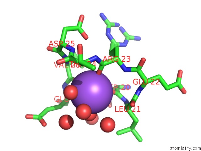
Sodium binding site 3 out of 4 in 5ur0
Go back to
Sodium binding site 3 out
of 4 in the Crystallographic Structure of Glyceraldehyde-3-Phosphate Dehydrogenase From Naegleria Gruberi
Mono view
Stereo pair view
Mono view
Stereo pair view
A full contact list of Sodium with other atoms in the Na binding
site number 3 of Crystallographic Structure of Glyceraldehyde-3-Phosphate Dehydrogenase From Naegleria Gruberi within 5.0Å range:
|
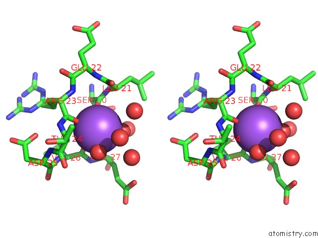
Sodium binding site 4 out of 4 in 5ur0
Go back to
Sodium binding site 4 out
of 4 in the Crystallographic Structure of Glyceraldehyde-3-Phosphate Dehydrogenase From Naegleria Gruberi
Mono view
Stereo pair view
Mono view
Stereo pair view
A full contact list of Sodium with other atoms in the Na binding
site number 4 of Crystallographic Structure of Glyceraldehyde-3-Phosphate Dehydrogenase From Naegleria Gruberi within 5.0Å range:
|
Reference:
A.T.P.Machado,
M.Silva,
J.Iulek.
Structural Studies of Glyceraldehyde-3-Phosphate Dehydrogenase From Naegleria Gruberi, the First One From Phylum Percolozoa. Biochim. Biophys. Acta V.1866 581 2018.
ISSN: ISSN 0006-3002
PubMed: 29501559
DOI: 10.1016/J.BBAPAP.2018.02.006
Page generated: Tue Oct 8 00:38:58 2024
ISSN: ISSN 0006-3002
PubMed: 29501559
DOI: 10.1016/J.BBAPAP.2018.02.006
Last articles
K in 4ZACK in 4ZND
K in 4ZAY
K in 4ZAN
K in 4ZAF
K in 4ZAG
K in 4ZAD
K in 4ZAB
K in 4ZAA
K in 4ZA9