Sodium »
PDB 5u6q-5uju »
5u92 »
Sodium in PDB 5u92: Crystal Structure of Arginine Kinase From the Spider Polybetes Pythagoricus in Complex with Arginine
Enzymatic activity of Crystal Structure of Arginine Kinase From the Spider Polybetes Pythagoricus in Complex with Arginine
All present enzymatic activity of Crystal Structure of Arginine Kinase From the Spider Polybetes Pythagoricus in Complex with Arginine:
2.7.3.3;
2.7.3.3;
Protein crystallography data
The structure of Crystal Structure of Arginine Kinase From the Spider Polybetes Pythagoricus in Complex with Arginine, PDB code: 5u92
was solved by
A.A.Lopez-Zavala,
C.F.Garcia,
J.Paredes-Hernandez,
V.Stojanoff,
R.R.Sotelo-Mundo,
with X-Ray Crystallography technique. A brief refinement statistics is given in the table below:
| Resolution Low / High (Å) | 19.73 / 2.00 |
| Space group | P 1 21 1 |
| Cell size a, b, c (Å), α, β, γ (°) | 48.090, 61.450, 60.600, 90.00, 95.75, 90.00 |
| R / Rfree (%) | 16.8 / 23.7 |
Sodium Binding Sites:
The binding sites of Sodium atom in the Crystal Structure of Arginine Kinase From the Spider Polybetes Pythagoricus in Complex with Arginine
(pdb code 5u92). This binding sites where shown within
5.0 Angstroms radius around Sodium atom.
In total 2 binding sites of Sodium where determined in the Crystal Structure of Arginine Kinase From the Spider Polybetes Pythagoricus in Complex with Arginine, PDB code: 5u92:
Jump to Sodium binding site number: 1; 2;
In total 2 binding sites of Sodium where determined in the Crystal Structure of Arginine Kinase From the Spider Polybetes Pythagoricus in Complex with Arginine, PDB code: 5u92:
Jump to Sodium binding site number: 1; 2;
Sodium binding site 1 out of 2 in 5u92
Go back to
Sodium binding site 1 out
of 2 in the Crystal Structure of Arginine Kinase From the Spider Polybetes Pythagoricus in Complex with Arginine
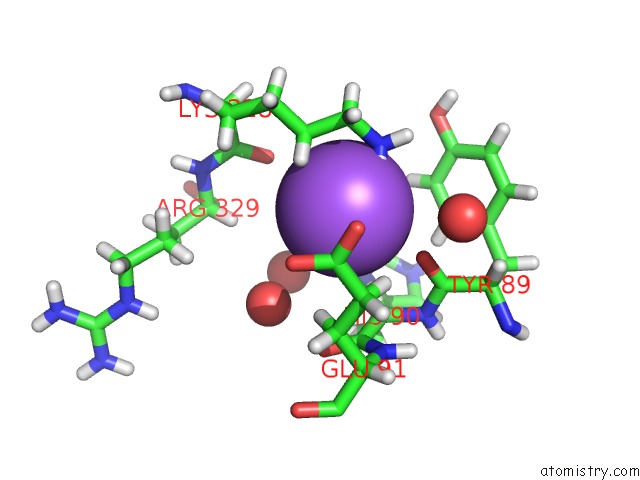
Mono view
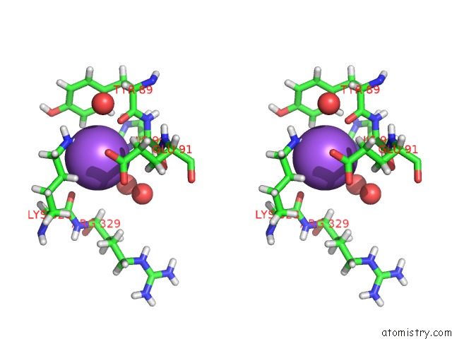
Stereo pair view
Mono view
Stereo pair view
A full contact list of Sodium with other atoms in the Na binding
site number 1 of Crystal Structure of Arginine Kinase From the Spider Polybetes Pythagoricus in Complex with Arginine within 5.0Å range:
|
Sodium binding site 2 out of 2 in 5u92
Go back to
Sodium binding site 2 out
of 2 in the Crystal Structure of Arginine Kinase From the Spider Polybetes Pythagoricus in Complex with Arginine
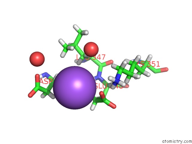
Mono view
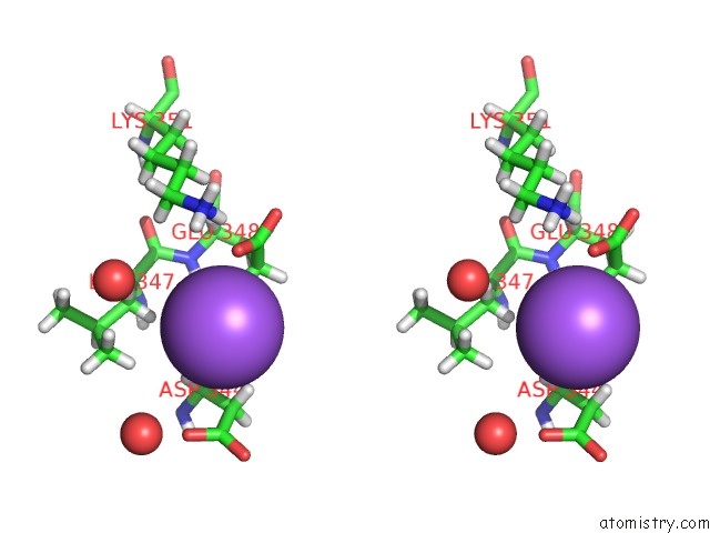
Stereo pair view
Mono view
Stereo pair view
A full contact list of Sodium with other atoms in the Na binding
site number 2 of Crystal Structure of Arginine Kinase From the Spider Polybetes Pythagoricus in Complex with Arginine within 5.0Å range:
|
Reference:
A.Laino,
A.A.Lopez-Zavala,
K.D.Garcia-Orozco,
J.S.Carrasco-Miranda,
M.Santana,
V.Stojanoff,
R.R.Sotelo-Mundo,
C.F.Garcia.
Biochemical and Structural Characterization of A Novel Arginine Kinase From the Spider Polybetes Pythagoricus. Peerj V. 5 E3787 2017.
ISSN: ESSN 2167-8359
PubMed: 28924503
DOI: 10.7717/PEERJ.3787
Page generated: Tue Oct 8 00:27:04 2024
ISSN: ESSN 2167-8359
PubMed: 28924503
DOI: 10.7717/PEERJ.3787
Last articles
Cl in 5FGACl in 5FG7
Cl in 5FF3
Cl in 5FF4
Cl in 5FDR
Cl in 5FF0
Cl in 5FF2
Cl in 5FEX
Cl in 5FEW
Cl in 5FEZ