Sodium »
PDB 5syu-5tde »
5t25 »
Sodium in PDB 5t25: Kinetic, Spectral and Structural Characterization of the Slow Binding Inhibitor Acetopyruvate with Dihydrodipicolinate Synthase From Escherichia Coli.
Enzymatic activity of Kinetic, Spectral and Structural Characterization of the Slow Binding Inhibitor Acetopyruvate with Dihydrodipicolinate Synthase From Escherichia Coli.
All present enzymatic activity of Kinetic, Spectral and Structural Characterization of the Slow Binding Inhibitor Acetopyruvate with Dihydrodipicolinate Synthase From Escherichia Coli.:
4.3.3.7;
4.3.3.7;
Protein crystallography data
The structure of Kinetic, Spectral and Structural Characterization of the Slow Binding Inhibitor Acetopyruvate with Dihydrodipicolinate Synthase From Escherichia Coli., PDB code: 5t25
was solved by
L.Chooback,
L.M.Thomas,
W.E.Karsten,
C.D.Fleming,
P.Seabourn,
with X-Ray Crystallography technique. A brief refinement statistics is given in the table below:
| Resolution Low / High (Å) | 26.81 / 1.99 |
| Space group | C 1 2 1 |
| Cell size a, b, c (Å), α, β, γ (°) | 136.647, 56.385, 101.530, 90.00, 127.62, 90.00 |
| R / Rfree (%) | 15.8 / 21.1 |
Sodium Binding Sites:
The binding sites of Sodium atom in the Kinetic, Spectral and Structural Characterization of the Slow Binding Inhibitor Acetopyruvate with Dihydrodipicolinate Synthase From Escherichia Coli.
(pdb code 5t25). This binding sites where shown within
5.0 Angstroms radius around Sodium atom.
In total 10 binding sites of Sodium where determined in the Kinetic, Spectral and Structural Characterization of the Slow Binding Inhibitor Acetopyruvate with Dihydrodipicolinate Synthase From Escherichia Coli., PDB code: 5t25:
Jump to Sodium binding site number: 1; 2; 3; 4; 5; 6; 7; 8; 9; 10;
In total 10 binding sites of Sodium where determined in the Kinetic, Spectral and Structural Characterization of the Slow Binding Inhibitor Acetopyruvate with Dihydrodipicolinate Synthase From Escherichia Coli., PDB code: 5t25:
Jump to Sodium binding site number: 1; 2; 3; 4; 5; 6; 7; 8; 9; 10;
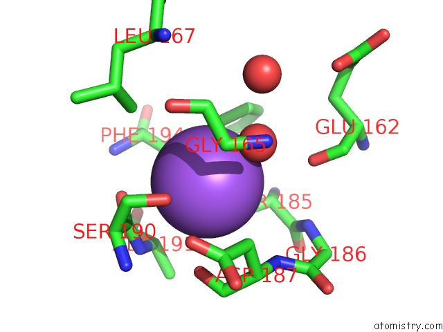
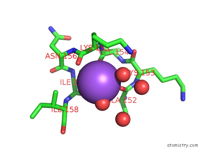
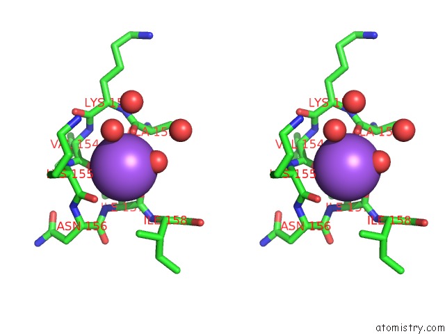
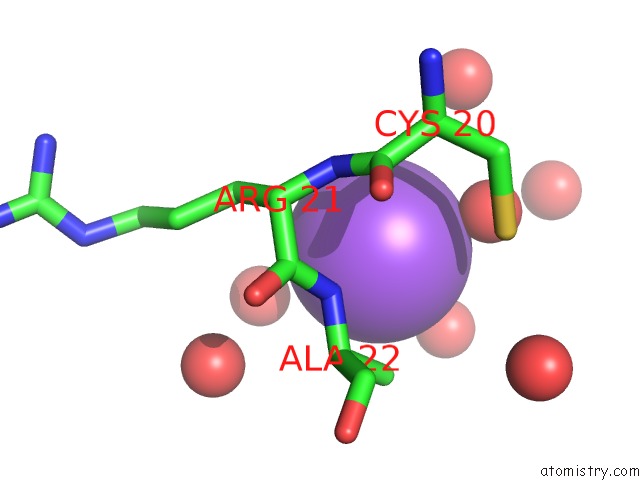
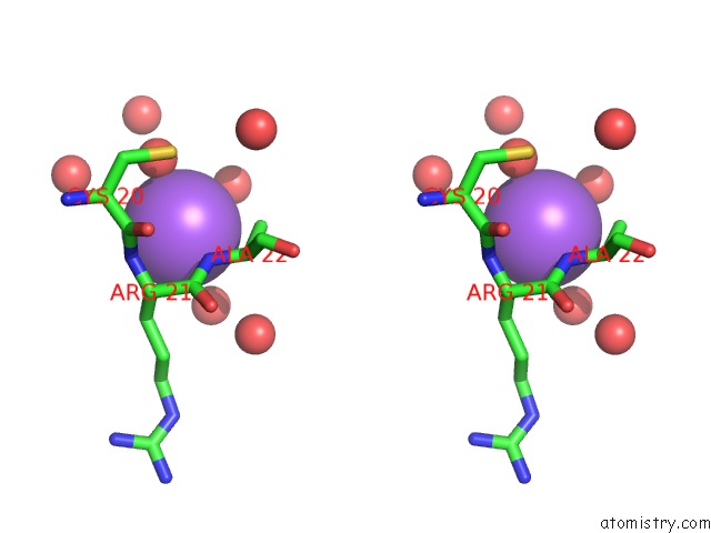
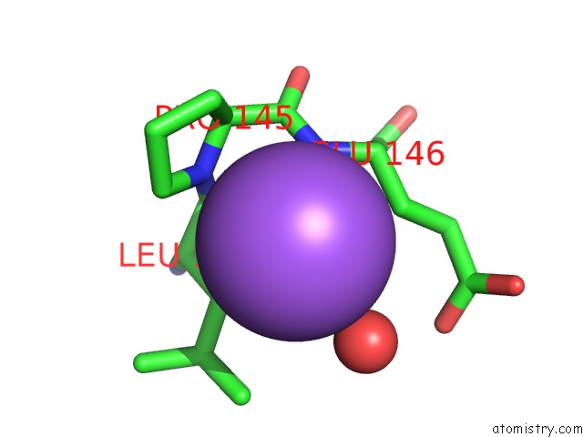
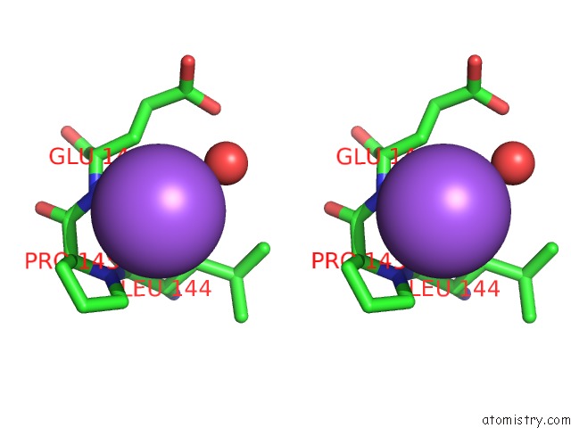
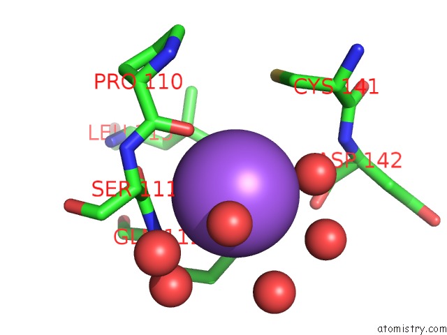
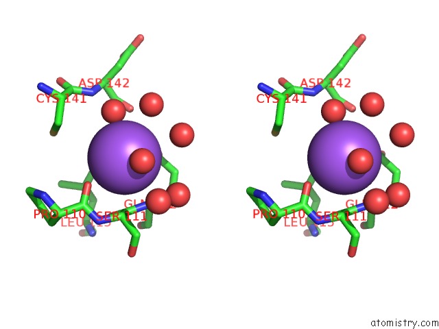
Sodium binding site 1 out of 10 in 5t25
Go back to
Sodium binding site 1 out
of 10 in the Kinetic, Spectral and Structural Characterization of the Slow Binding Inhibitor Acetopyruvate with Dihydrodipicolinate Synthase From Escherichia Coli.
Mono view
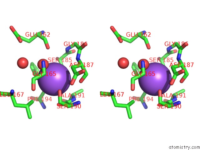
Stereo pair view
Mono view
Stereo pair view
A full contact list of Sodium with other atoms in the Na binding
site number 1 of Kinetic, Spectral and Structural Characterization of the Slow Binding Inhibitor Acetopyruvate with Dihydrodipicolinate Synthase From Escherichia Coli. within 5.0Å range:
|
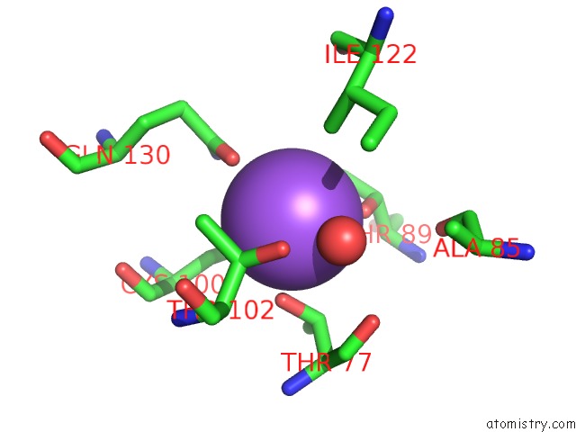
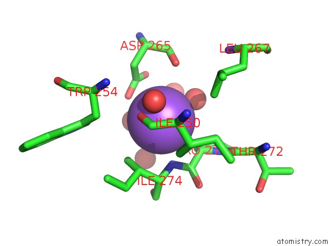
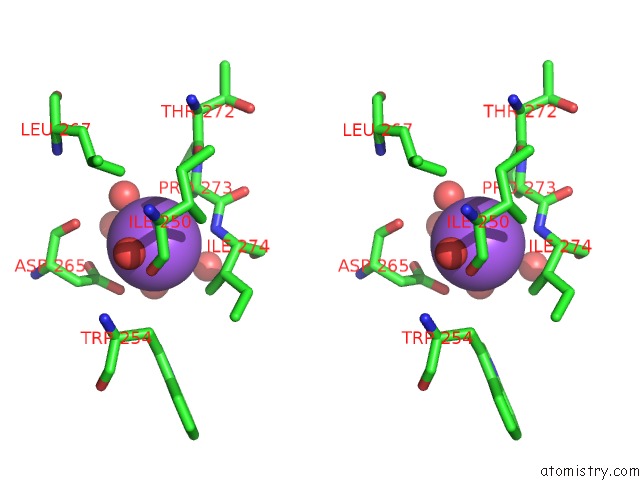
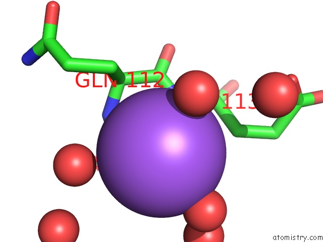
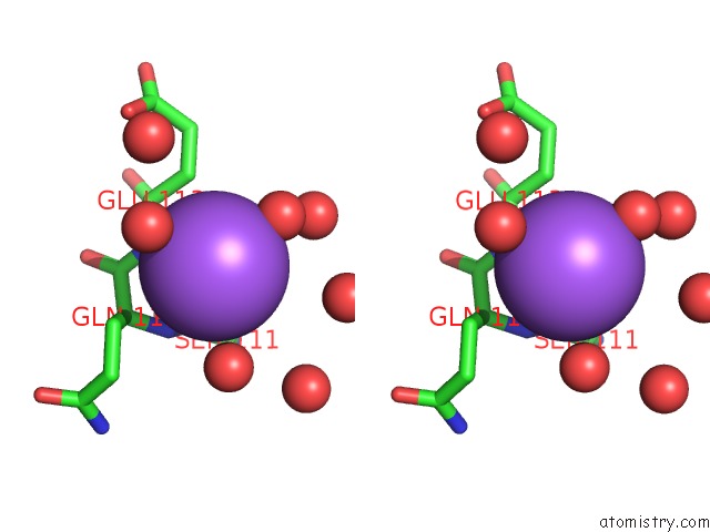
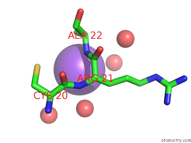
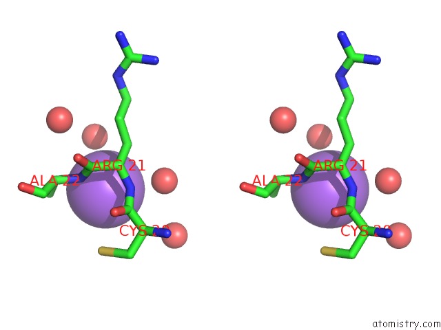
Sodium binding site 2 out of 10 in 5t25
Go back to
Sodium binding site 2 out
of 10 in the Kinetic, Spectral and Structural Characterization of the Slow Binding Inhibitor Acetopyruvate with Dihydrodipicolinate Synthase From Escherichia Coli.
Mono view
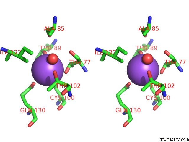
Stereo pair view
Mono view
Stereo pair view
A full contact list of Sodium with other atoms in the Na binding
site number 2 of Kinetic, Spectral and Structural Characterization of the Slow Binding Inhibitor Acetopyruvate with Dihydrodipicolinate Synthase From Escherichia Coli. within 5.0Å range:
|
Sodium binding site 3 out of 10 in 5t25
Go back to
Sodium binding site 3 out
of 10 in the Kinetic, Spectral and Structural Characterization of the Slow Binding Inhibitor Acetopyruvate with Dihydrodipicolinate Synthase From Escherichia Coli.
Mono view
Stereo pair view
Mono view
Stereo pair view
A full contact list of Sodium with other atoms in the Na binding
site number 3 of Kinetic, Spectral and Structural Characterization of the Slow Binding Inhibitor Acetopyruvate with Dihydrodipicolinate Synthase From Escherichia Coli. within 5.0Å range:
|
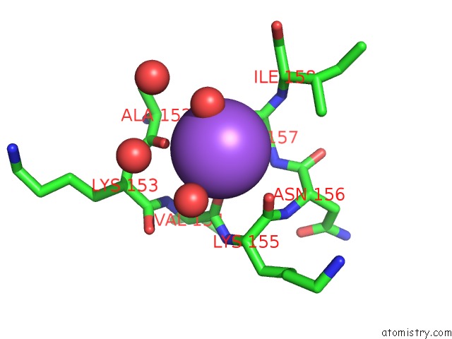
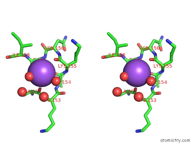
Sodium binding site 4 out of 10 in 5t25
Go back to
Sodium binding site 4 out
of 10 in the Kinetic, Spectral and Structural Characterization of the Slow Binding Inhibitor Acetopyruvate with Dihydrodipicolinate Synthase From Escherichia Coli.
Mono view
Stereo pair view
Mono view
Stereo pair view
A full contact list of Sodium with other atoms in the Na binding
site number 4 of Kinetic, Spectral and Structural Characterization of the Slow Binding Inhibitor Acetopyruvate with Dihydrodipicolinate Synthase From Escherichia Coli. within 5.0Å range:
|
Sodium binding site 5 out of 10 in 5t25
Go back to
Sodium binding site 5 out
of 10 in the Kinetic, Spectral and Structural Characterization of the Slow Binding Inhibitor Acetopyruvate with Dihydrodipicolinate Synthase From Escherichia Coli.
Mono view
Stereo pair view
Mono view
Stereo pair view
A full contact list of Sodium with other atoms in the Na binding
site number 5 of Kinetic, Spectral and Structural Characterization of the Slow Binding Inhibitor Acetopyruvate with Dihydrodipicolinate Synthase From Escherichia Coli. within 5.0Å range:
|
Sodium binding site 6 out of 10 in 5t25
Go back to
Sodium binding site 6 out
of 10 in the Kinetic, Spectral and Structural Characterization of the Slow Binding Inhibitor Acetopyruvate with Dihydrodipicolinate Synthase From Escherichia Coli.
Mono view
Stereo pair view
Mono view
Stereo pair view
A full contact list of Sodium with other atoms in the Na binding
site number 6 of Kinetic, Spectral and Structural Characterization of the Slow Binding Inhibitor Acetopyruvate with Dihydrodipicolinate Synthase From Escherichia Coli. within 5.0Å range:
|
Sodium binding site 7 out of 10 in 5t25
Go back to
Sodium binding site 7 out
of 10 in the Kinetic, Spectral and Structural Characterization of the Slow Binding Inhibitor Acetopyruvate with Dihydrodipicolinate Synthase From Escherichia Coli.
Mono view
Stereo pair view
Mono view
Stereo pair view
A full contact list of Sodium with other atoms in the Na binding
site number 7 of Kinetic, Spectral and Structural Characterization of the Slow Binding Inhibitor Acetopyruvate with Dihydrodipicolinate Synthase From Escherichia Coli. within 5.0Å range:
|
Sodium binding site 8 out of 10 in 5t25
Go back to
Sodium binding site 8 out
of 10 in the Kinetic, Spectral and Structural Characterization of the Slow Binding Inhibitor Acetopyruvate with Dihydrodipicolinate Synthase From Escherichia Coli.
Mono view
Stereo pair view
Mono view
Stereo pair view
A full contact list of Sodium with other atoms in the Na binding
site number 8 of Kinetic, Spectral and Structural Characterization of the Slow Binding Inhibitor Acetopyruvate with Dihydrodipicolinate Synthase From Escherichia Coli. within 5.0Å range:
|
Sodium binding site 9 out of 10 in 5t25
Go back to
Sodium binding site 9 out
of 10 in the Kinetic, Spectral and Structural Characterization of the Slow Binding Inhibitor Acetopyruvate with Dihydrodipicolinate Synthase From Escherichia Coli.
Mono view
Stereo pair view
Mono view
Stereo pair view
A full contact list of Sodium with other atoms in the Na binding
site number 9 of Kinetic, Spectral and Structural Characterization of the Slow Binding Inhibitor Acetopyruvate with Dihydrodipicolinate Synthase From Escherichia Coli. within 5.0Å range:
|
Sodium binding site 10 out of 10 in 5t25
Go back to
Sodium binding site 10 out
of 10 in the Kinetic, Spectral and Structural Characterization of the Slow Binding Inhibitor Acetopyruvate with Dihydrodipicolinate Synthase From Escherichia Coli.
Mono view
Stereo pair view
Mono view
Stereo pair view
A full contact list of Sodium with other atoms in the Na binding
site number 10 of Kinetic, Spectral and Structural Characterization of the Slow Binding Inhibitor Acetopyruvate with Dihydrodipicolinate Synthase From Escherichia Coli. within 5.0Å range:
|
Reference:
L.Chooback,
L.M.Thomas,
W.E.Karsten,
C.D.Fleming,
P.Seabourn.
Kinetic, Spectral and Structural Characterization of the Slow Binding Inhibitor Acetopyruvate with Dihydrodipicolinate Synthase From Escherichia Coli. To Be Published.
Page generated: Mon Aug 18 02:10:51 2025
Last articles
Zn in 1XJSZn in 1XFO
Zn in 1XJO
Zn in 1XJH
Zn in 1XI2
Zn in 1XGE
Zn in 1XB0
Zn in 1XB1
Zn in 1XEV
Zn in 1XF7