Sodium »
PDB 5rdn-5syl »
5sus »
Sodium in PDB 5sus: X-Ray Crystallographic Structure of A Covalent Trimer Derived From A- Beta 17_36. X-Ray Diffractometer Data Set. (Orn)Cvf(Mea)Ced(Orn) Aiigl(Orn)V.
Protein crystallography data
The structure of X-Ray Crystallographic Structure of A Covalent Trimer Derived From A- Beta 17_36. X-Ray Diffractometer Data Set. (Orn)Cvf(Mea)Ced(Orn) Aiigl(Orn)V., PDB code: 5sus
was solved by
A.G.Kreutzer,
S.Yoo,
J.S.Nowick,
with X-Ray Crystallography technique. A brief refinement statistics is given in the table below:
| Resolution Low / High (Å) | 26.40 / 2.35 |
| Space group | P 63 2 2 |
| Cell size a, b, c (Å), α, β, γ (°) | 57.085, 57.085, 93.682, 90.00, 90.00, 120.00 |
| R / Rfree (%) | 21.9 / 27.9 |
Other elements in 5sus:
The structure of X-Ray Crystallographic Structure of A Covalent Trimer Derived From A- Beta 17_36. X-Ray Diffractometer Data Set. (Orn)Cvf(Mea)Ced(Orn) Aiigl(Orn)V. also contains other interesting chemical elements:
| Chlorine | (Cl) | 1 atom |
Sodium Binding Sites:
The binding sites of Sodium atom in the X-Ray Crystallographic Structure of A Covalent Trimer Derived From A- Beta 17_36. X-Ray Diffractometer Data Set. (Orn)Cvf(Mea)Ced(Orn) Aiigl(Orn)V.
(pdb code 5sus). This binding sites where shown within
5.0 Angstroms radius around Sodium atom.
In total only one binding site of Sodium was determined in the X-Ray Crystallographic Structure of A Covalent Trimer Derived From A- Beta 17_36. X-Ray Diffractometer Data Set. (Orn)Cvf(Mea)Ced(Orn) Aiigl(Orn)V., PDB code: 5sus:
In total only one binding site of Sodium was determined in the X-Ray Crystallographic Structure of A Covalent Trimer Derived From A- Beta 17_36. X-Ray Diffractometer Data Set. (Orn)Cvf(Mea)Ced(Orn) Aiigl(Orn)V., PDB code: 5sus:
Sodium binding site 1 out of 1 in 5sus
Go back to
Sodium binding site 1 out
of 1 in the X-Ray Crystallographic Structure of A Covalent Trimer Derived From A- Beta 17_36. X-Ray Diffractometer Data Set. (Orn)Cvf(Mea)Ced(Orn) Aiigl(Orn)V.
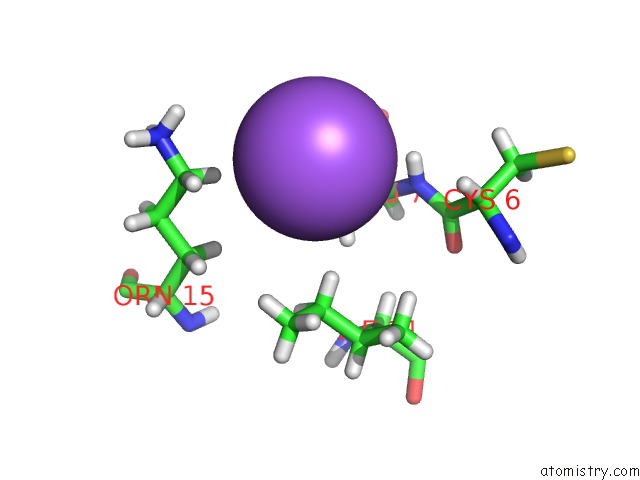
Mono view
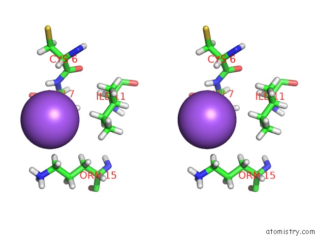
Stereo pair view
Mono view
Stereo pair view
A full contact list of Sodium with other atoms in the Na binding
site number 1 of X-Ray Crystallographic Structure of A Covalent Trimer Derived From A- Beta 17_36. X-Ray Diffractometer Data Set. (Orn)Cvf(Mea)Ced(Orn) Aiigl(Orn)V. within 5.0Å range:
|
Reference:
A.G.Kreutzer,
S.Yoo,
R.K.Spencer,
J.S.Nowick.
X-Ray Crystallographic Structure of A Covalent Trimer Derived From A-Beta 17_36. X-Ray Diffractometer Data Set. (Orn)Cvf(Mea)Ced(Orn)Aiigl(Orn)V. To Be Published.
Page generated: Mon Oct 7 23:54:32 2024
Last articles
Mg in 5DS5Mg in 5DRZ
Mg in 5DRI
Mg in 5DRC
Mg in 5DRD
Mg in 5DQL
Mg in 5DR2
Mg in 5DQZ
Mg in 5DQK
Mg in 5DOU