Sodium »
PDB 5l6q-5lq6 »
5lhv »
Sodium in PDB 5lhv: X-Ray Structure of Uridine Phosphorylase From Vibrio Cholerae in Complex with Uridine and Sulfate Ion at 1.29 A Resolution
Enzymatic activity of X-Ray Structure of Uridine Phosphorylase From Vibrio Cholerae in Complex with Uridine and Sulfate Ion at 1.29 A Resolution
All present enzymatic activity of X-Ray Structure of Uridine Phosphorylase From Vibrio Cholerae in Complex with Uridine and Sulfate Ion at 1.29 A Resolution:
2.4.2.3;
2.4.2.3;
Protein crystallography data
The structure of X-Ray Structure of Uridine Phosphorylase From Vibrio Cholerae in Complex with Uridine and Sulfate Ion at 1.29 A Resolution, PDB code: 5lhv
was solved by
I.I.Prokofev,
A.A.Lashkov,
A.G.Gabdoulkhakov,
V.V.Balaev,
C.Betzel,
A.M.Mikhailov,
with X-Ray Crystallography technique. A brief refinement statistics is given in the table below:
| Resolution Low / High (Å) | 33.57 / 1.29 |
| Space group | P 1 |
| Cell size a, b, c (Å), α, β, γ (°) | 64.223, 71.648, 88.878, 69.70, 72.70, 86.24 |
| R / Rfree (%) | 14.5 / 17.8 |
Other elements in 5lhv:
The structure of X-Ray Structure of Uridine Phosphorylase From Vibrio Cholerae in Complex with Uridine and Sulfate Ion at 1.29 A Resolution also contains other interesting chemical elements:
| Magnesium | (Mg) | 7 atoms |
| Chlorine | (Cl) | 6 atoms |
Sodium Binding Sites:
The binding sites of Sodium atom in the X-Ray Structure of Uridine Phosphorylase From Vibrio Cholerae in Complex with Uridine and Sulfate Ion at 1.29 A Resolution
(pdb code 5lhv). This binding sites where shown within
5.0 Angstroms radius around Sodium atom.
In total 3 binding sites of Sodium where determined in the X-Ray Structure of Uridine Phosphorylase From Vibrio Cholerae in Complex with Uridine and Sulfate Ion at 1.29 A Resolution, PDB code: 5lhv:
Jump to Sodium binding site number: 1; 2; 3;
In total 3 binding sites of Sodium where determined in the X-Ray Structure of Uridine Phosphorylase From Vibrio Cholerae in Complex with Uridine and Sulfate Ion at 1.29 A Resolution, PDB code: 5lhv:
Jump to Sodium binding site number: 1; 2; 3;
Sodium binding site 1 out of 3 in 5lhv
Go back to
Sodium binding site 1 out
of 3 in the X-Ray Structure of Uridine Phosphorylase From Vibrio Cholerae in Complex with Uridine and Sulfate Ion at 1.29 A Resolution

Mono view
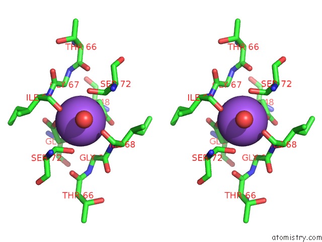
Stereo pair view
Mono view
Stereo pair view
A full contact list of Sodium with other atoms in the Na binding
site number 1 of X-Ray Structure of Uridine Phosphorylase From Vibrio Cholerae in Complex with Uridine and Sulfate Ion at 1.29 A Resolution within 5.0Å range:
|
Sodium binding site 2 out of 3 in 5lhv
Go back to
Sodium binding site 2 out
of 3 in the X-Ray Structure of Uridine Phosphorylase From Vibrio Cholerae in Complex with Uridine and Sulfate Ion at 1.29 A Resolution

Mono view

Stereo pair view
Mono view
Stereo pair view
A full contact list of Sodium with other atoms in the Na binding
site number 2 of X-Ray Structure of Uridine Phosphorylase From Vibrio Cholerae in Complex with Uridine and Sulfate Ion at 1.29 A Resolution within 5.0Å range:
|
Sodium binding site 3 out of 3 in 5lhv
Go back to
Sodium binding site 3 out
of 3 in the X-Ray Structure of Uridine Phosphorylase From Vibrio Cholerae in Complex with Uridine and Sulfate Ion at 1.29 A Resolution

Mono view

Stereo pair view
Mono view
Stereo pair view
A full contact list of Sodium with other atoms in the Na binding
site number 3 of X-Ray Structure of Uridine Phosphorylase From Vibrio Cholerae in Complex with Uridine and Sulfate Ion at 1.29 A Resolution within 5.0Å range:
|
Reference:
I.I.Prokofev,
A.A.Lashkov,
A.G.Gabdoulkhakov,
V.V.Balaev,
C.Betzel,
A.M.Mikhailov.
X-Ray Structure of Uridine Phosphorylase From Vibrio Cholerae in Complex with Uridine and Sulfate Ion at 1.29 A Resolution To Be Published.
Page generated: Mon Oct 7 22:21:04 2024
Last articles
I in 4JIJI in 4JCY
I in 4J3X
I in 4IQZ
I in 4J3W
I in 4J3V
I in 4IZG
I in 4J3T
I in 4J3S
I in 4J2V