Sodium »
PDB 5j2d-5jim »
5jgl »
Sodium in PDB 5jgl: Crystal Structure of Gtma in Complex with S-Adenosylmethionine
Protein crystallography data
The structure of Crystal Structure of Gtma in Complex with S-Adenosylmethionine, PDB code: 5jgl
was solved by
S.K.Dolan,
T.Bock,
V.Hering,
G.W.Jones,
W.Blankenfeldt,
S.Doyle,
with X-Ray Crystallography technique. A brief refinement statistics is given in the table below:
| Resolution Low / High (Å) | 49.28 / 2.28 |
| Space group | P 1 21 1 |
| Cell size a, b, c (Å), α, β, γ (°) | 45.989, 114.142, 56.438, 90.00, 104.55, 90.00 |
| R / Rfree (%) | 19.2 / 23.4 |
Sodium Binding Sites:
The binding sites of Sodium atom in the Crystal Structure of Gtma in Complex with S-Adenosylmethionine
(pdb code 5jgl). This binding sites where shown within
5.0 Angstroms radius around Sodium atom.
In total 2 binding sites of Sodium where determined in the Crystal Structure of Gtma in Complex with S-Adenosylmethionine, PDB code: 5jgl:
Jump to Sodium binding site number: 1; 2;
In total 2 binding sites of Sodium where determined in the Crystal Structure of Gtma in Complex with S-Adenosylmethionine, PDB code: 5jgl:
Jump to Sodium binding site number: 1; 2;
Sodium binding site 1 out of 2 in 5jgl
Go back to
Sodium binding site 1 out
of 2 in the Crystal Structure of Gtma in Complex with S-Adenosylmethionine
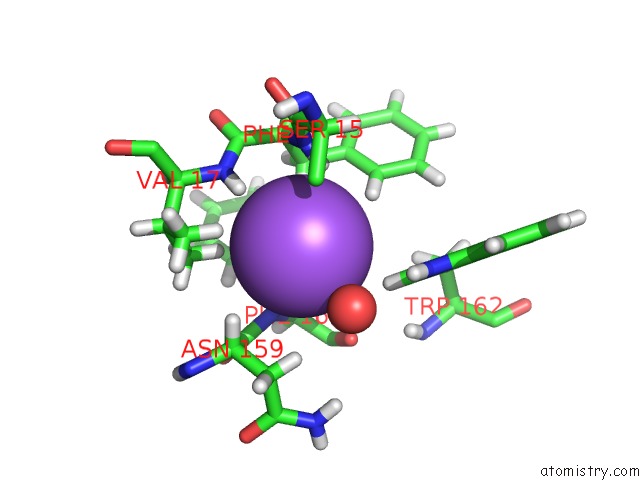
Mono view
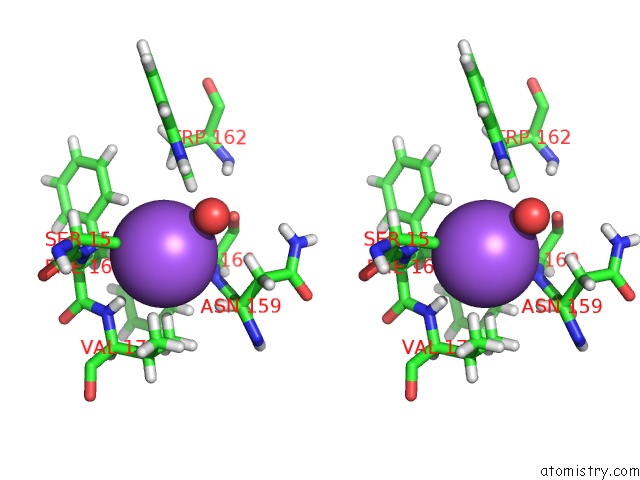
Stereo pair view
Mono view
Stereo pair view
A full contact list of Sodium with other atoms in the Na binding
site number 1 of Crystal Structure of Gtma in Complex with S-Adenosylmethionine within 5.0Å range:
|
Sodium binding site 2 out of 2 in 5jgl
Go back to
Sodium binding site 2 out
of 2 in the Crystal Structure of Gtma in Complex with S-Adenosylmethionine
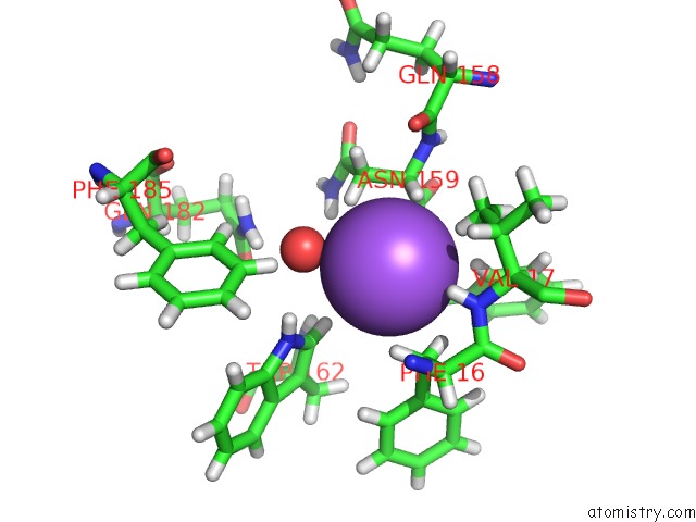
Mono view
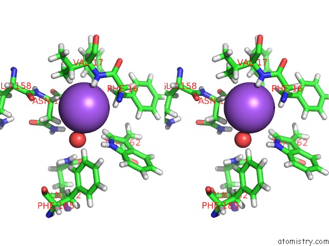
Stereo pair view
Mono view
Stereo pair view
A full contact list of Sodium with other atoms in the Na binding
site number 2 of Crystal Structure of Gtma in Complex with S-Adenosylmethionine within 5.0Å range:
|
Reference:
S.K.Dolan,
T.Bock,
V.Hering,
R.A.Owens,
G.W.Jones,
W.Blankenfeldt,
S.Doyle.
Structural, Mechanistic and Functional Insight Into Gliotoxinbis-Thiomethylation Inaspergillus Fumigatus. Open Biol V. 7 2017.
ISSN: ESSN 2046-2441
PubMed: 28179499
DOI: 10.1098/RSOB.160292
Page generated: Mon Aug 18 00:30:42 2025
ISSN: ESSN 2046-2441
PubMed: 28179499
DOI: 10.1098/RSOB.160292
Last articles
Na in 7M4FNa in 7M4C
Na in 7M4E
Na in 7M4D
Na in 7M4B
Na in 7M48
Na in 7M4A
Na in 7M49
Na in 7M47
Na in 7M46