Sodium »
PDB 5gwl-5hn2 »
5h9v »
Sodium in PDB 5h9v: Crystal Structure of Regnase Pin Domain, Form I
Protein crystallography data
The structure of Crystal Structure of Regnase Pin Domain, Form I, PDB code: 5h9v
was solved by
M.Yokogawa,
T.Tsushima,
W.Adachi,
N.N.Noda,
F.Inagaki,
with X-Ray Crystallography technique. A brief refinement statistics is given in the table below:
| Resolution Low / High (Å) | 47.49 / 2.75 |
| Space group | P 32 2 1 |
| Cell size a, b, c (Å), α, β, γ (°) | 113.370, 113.370, 187.438, 90.00, 90.00, 120.00 |
| R / Rfree (%) | 18.9 / 22.6 |
Sodium Binding Sites:
The binding sites of Sodium atom in the Crystal Structure of Regnase Pin Domain, Form I
(pdb code 5h9v). This binding sites where shown within
5.0 Angstroms radius around Sodium atom.
In total 4 binding sites of Sodium where determined in the Crystal Structure of Regnase Pin Domain, Form I, PDB code: 5h9v:
Jump to Sodium binding site number: 1; 2; 3; 4;
In total 4 binding sites of Sodium where determined in the Crystal Structure of Regnase Pin Domain, Form I, PDB code: 5h9v:
Jump to Sodium binding site number: 1; 2; 3; 4;
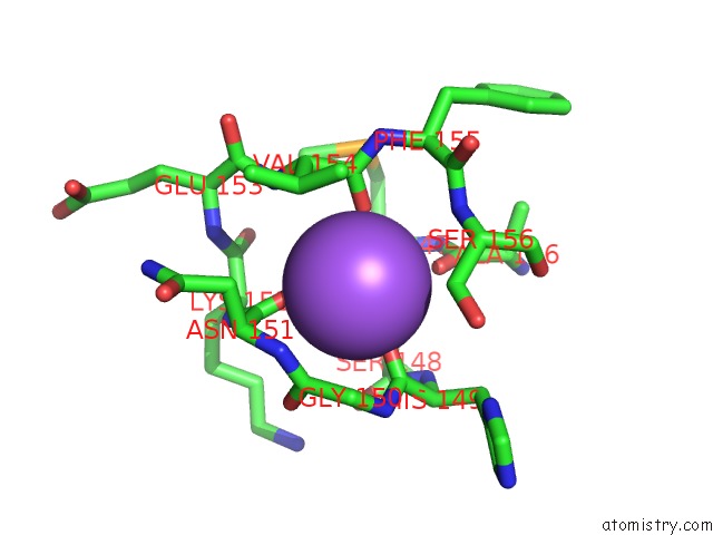
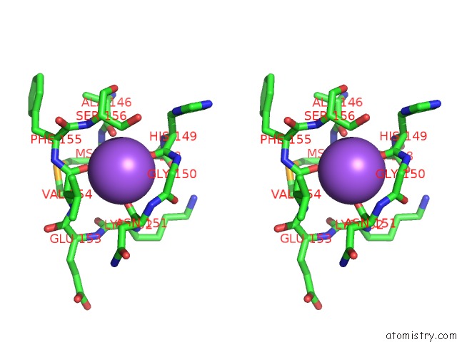
Sodium binding site 1 out of 4 in 5h9v
Go back to
Sodium binding site 1 out
of 4 in the Crystal Structure of Regnase Pin Domain, Form I
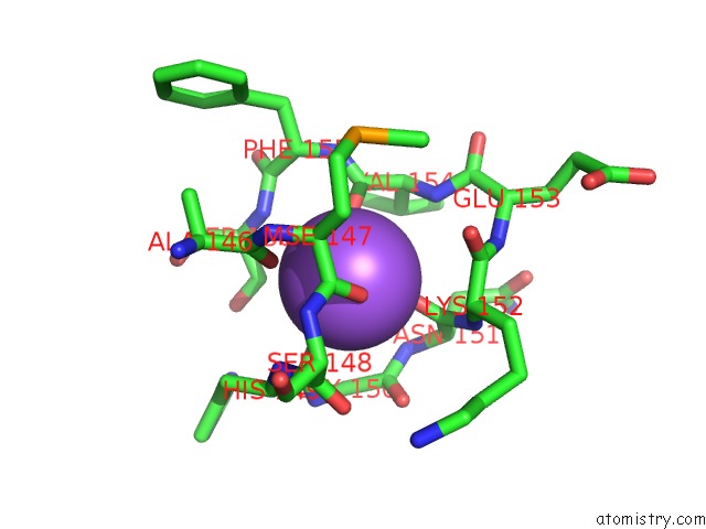
Mono view
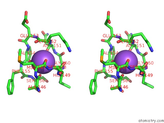
Stereo pair view
Mono view
Stereo pair view
A full contact list of Sodium with other atoms in the Na binding
site number 1 of Crystal Structure of Regnase Pin Domain, Form I within 5.0Å range:
|
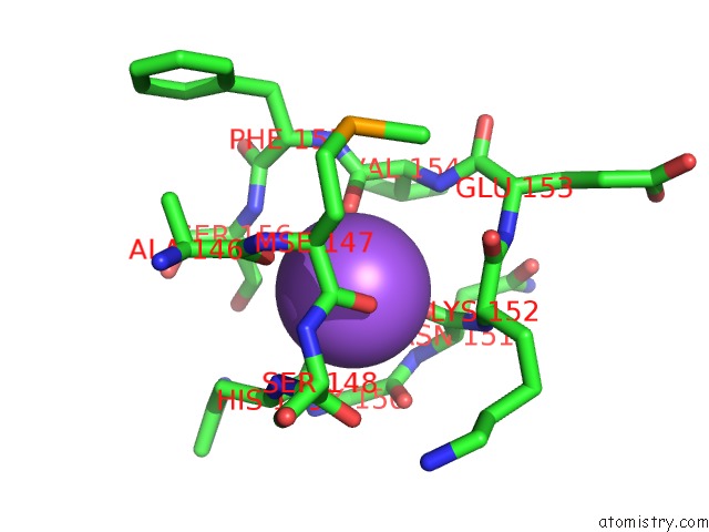
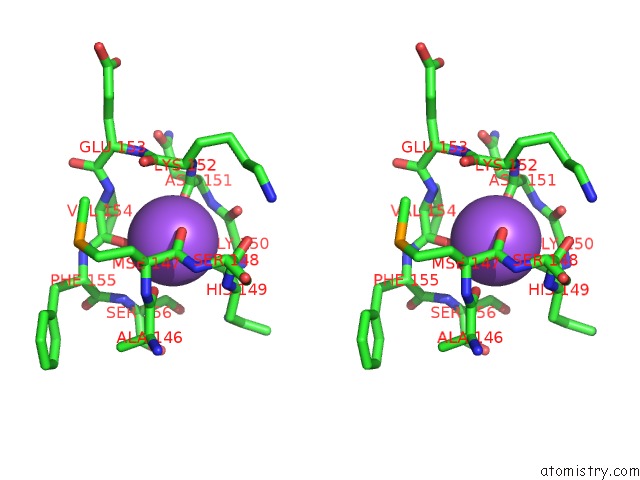
Sodium binding site 2 out of 4 in 5h9v
Go back to
Sodium binding site 2 out
of 4 in the Crystal Structure of Regnase Pin Domain, Form I
Mono view
Stereo pair view
Mono view
Stereo pair view
A full contact list of Sodium with other atoms in the Na binding
site number 2 of Crystal Structure of Regnase Pin Domain, Form I within 5.0Å range:
|
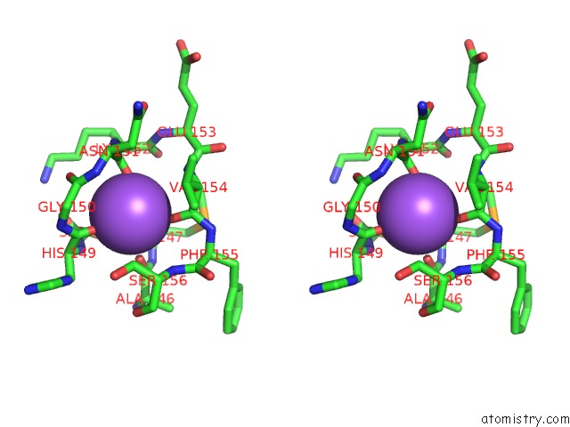
Sodium binding site 3 out of 4 in 5h9v
Go back to
Sodium binding site 3 out
of 4 in the Crystal Structure of Regnase Pin Domain, Form I
Mono view
Stereo pair view
Mono view
Stereo pair view
A full contact list of Sodium with other atoms in the Na binding
site number 3 of Crystal Structure of Regnase Pin Domain, Form I within 5.0Å range:
|
Sodium binding site 4 out of 4 in 5h9v
Go back to
Sodium binding site 4 out
of 4 in the Crystal Structure of Regnase Pin Domain, Form I
Mono view
Stereo pair view
Mono view
Stereo pair view
A full contact list of Sodium with other atoms in the Na binding
site number 4 of Crystal Structure of Regnase Pin Domain, Form I within 5.0Å range:
|
Reference:
M.Yokogawa,
T.Tsushima,
N.N.Noda,
H.Kumeta,
Y.Enokizono,
K.Yamashita,
D.M.Standley,
O.Takeuchi,
S.Akira,
F.Inagaki.
Structural Basis For the Regulation of Enzymatic Activity of Regnase-1 By Domain-Domain Interactions Sci Rep V. 6 22324 2016.
ISSN: ESSN 2045-2322
PubMed: 26927947
DOI: 10.1038/SREP22324
Page generated: Mon Oct 7 21:20:52 2024
ISSN: ESSN 2045-2322
PubMed: 26927947
DOI: 10.1038/SREP22324
Last articles
Mg in 4ZK5Mg in 4ZJJ
Mg in 4ZJI
Mg in 4ZK4
Mg in 4ZIR
Mg in 4ZIY
Mg in 4ZIB
Mg in 4ZI7
Mg in 4ZIL
Mg in 4ZI5