Sodium »
PDB 5fje-5g3f »
5fub »
Sodium in PDB 5fub: Crystal Structure of Zebrafish Protein Arginine Methyltransferase 2 Catalytic Domain with Sah
Enzymatic activity of Crystal Structure of Zebrafish Protein Arginine Methyltransferase 2 Catalytic Domain with Sah
All present enzymatic activity of Crystal Structure of Zebrafish Protein Arginine Methyltransferase 2 Catalytic Domain with Sah:
2.1.1.125;
2.1.1.125;
Protein crystallography data
The structure of Crystal Structure of Zebrafish Protein Arginine Methyltransferase 2 Catalytic Domain with Sah, PDB code: 5fub
was solved by
V.Cura,
N.Troffer-Charlier,
N.Marechal,
L.Bonnefond,
J.Cavarelli,
with X-Ray Crystallography technique. A brief refinement statistics is given in the table below:
| Resolution Low / High (Å) | 38.41 / 2.00 |
| Space group | H 3 2 |
| Cell size a, b, c (Å), α, β, γ (°) | 147.070, 147.070, 127.390, 90.00, 90.00, 120.00 |
| R / Rfree (%) | 18.2 / 21.8 |
Other elements in 5fub:
The structure of Crystal Structure of Zebrafish Protein Arginine Methyltransferase 2 Catalytic Domain with Sah also contains other interesting chemical elements:
| Chlorine | (Cl) | 1 atom |
Sodium Binding Sites:
The binding sites of Sodium atom in the Crystal Structure of Zebrafish Protein Arginine Methyltransferase 2 Catalytic Domain with Sah
(pdb code 5fub). This binding sites where shown within
5.0 Angstroms radius around Sodium atom.
In total 2 binding sites of Sodium where determined in the Crystal Structure of Zebrafish Protein Arginine Methyltransferase 2 Catalytic Domain with Sah, PDB code: 5fub:
Jump to Sodium binding site number: 1; 2;
In total 2 binding sites of Sodium where determined in the Crystal Structure of Zebrafish Protein Arginine Methyltransferase 2 Catalytic Domain with Sah, PDB code: 5fub:
Jump to Sodium binding site number: 1; 2;
Sodium binding site 1 out of 2 in 5fub
Go back to
Sodium binding site 1 out
of 2 in the Crystal Structure of Zebrafish Protein Arginine Methyltransferase 2 Catalytic Domain with Sah
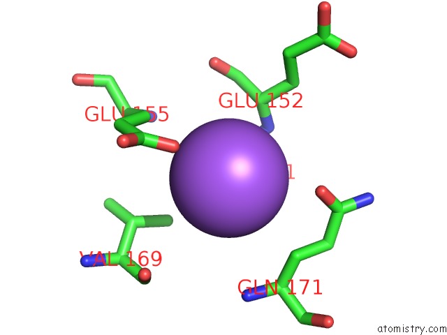
Mono view
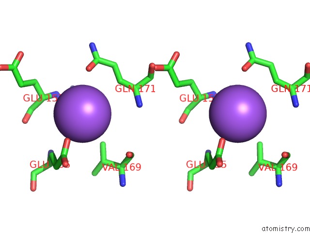
Stereo pair view
Mono view
Stereo pair view
A full contact list of Sodium with other atoms in the Na binding
site number 1 of Crystal Structure of Zebrafish Protein Arginine Methyltransferase 2 Catalytic Domain with Sah within 5.0Å range:
|
Sodium binding site 2 out of 2 in 5fub
Go back to
Sodium binding site 2 out
of 2 in the Crystal Structure of Zebrafish Protein Arginine Methyltransferase 2 Catalytic Domain with Sah
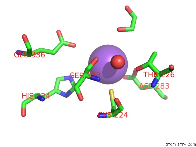
Mono view
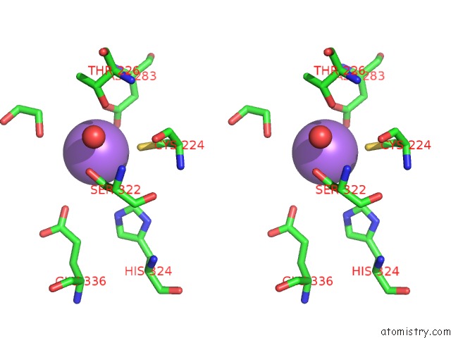
Stereo pair view
Mono view
Stereo pair view
A full contact list of Sodium with other atoms in the Na binding
site number 2 of Crystal Structure of Zebrafish Protein Arginine Methyltransferase 2 Catalytic Domain with Sah within 5.0Å range:
|
Reference:
V.Cura,
N.Marechal,
N.Troffer-Charlier,
J.M.Strub,
M.J.Van Haren,
N.I.Martin,
S.Cianferani,
L.Bonnefond,
J.Cavarelli.
Structural Studies of Protein Arginine Methyltransferase 2 Reveal Its Interactions with Potential Substrates and Inhibitors. Febs J. V. 284 77 2017.
ISSN: ISSN 1742-4658
PubMed: 27879050
DOI: 10.1111/FEBS.13953
Page generated: Mon Oct 7 21:06:01 2024
ISSN: ISSN 1742-4658
PubMed: 27879050
DOI: 10.1111/FEBS.13953
Last articles
Mg in 1S5LMg in 1S8F
Mg in 1S83
Mg in 1S77
Mg in 1S76
Mg in 1S4E
Mg in 1S6P
Mg in 1S6H
Mg in 1S5J
Mg in 1S5G