Sodium »
PDB 5ddq-5dy9 »
5dnu »
Sodium in PDB 5dnu: Crystal Structure of Striga KAI2-Like Protein in Complex with Karrikin
Protein crystallography data
The structure of Crystal Structure of Striga KAI2-Like Protein in Complex with Karrikin, PDB code: 5dnu
was solved by
Y.Xu,
T.Miyakawa,
A.Nakamura,
M.Tanokura,
with X-Ray Crystallography technique. A brief refinement statistics is given in the table below:
| Resolution Low / High (Å) | 19.70 / 1.20 |
| Space group | P 61 2 2 |
| Cell size a, b, c (Å), α, β, γ (°) | 75.757, 75.757, 181.266, 90.00, 90.00, 120.00 |
| R / Rfree (%) | 14 / 15.5 |
Sodium Binding Sites:
The binding sites of Sodium atom in the Crystal Structure of Striga KAI2-Like Protein in Complex with Karrikin
(pdb code 5dnu). This binding sites where shown within
5.0 Angstroms radius around Sodium atom.
In total 3 binding sites of Sodium where determined in the Crystal Structure of Striga KAI2-Like Protein in Complex with Karrikin, PDB code: 5dnu:
Jump to Sodium binding site number: 1; 2; 3;
In total 3 binding sites of Sodium where determined in the Crystal Structure of Striga KAI2-Like Protein in Complex with Karrikin, PDB code: 5dnu:
Jump to Sodium binding site number: 1; 2; 3;
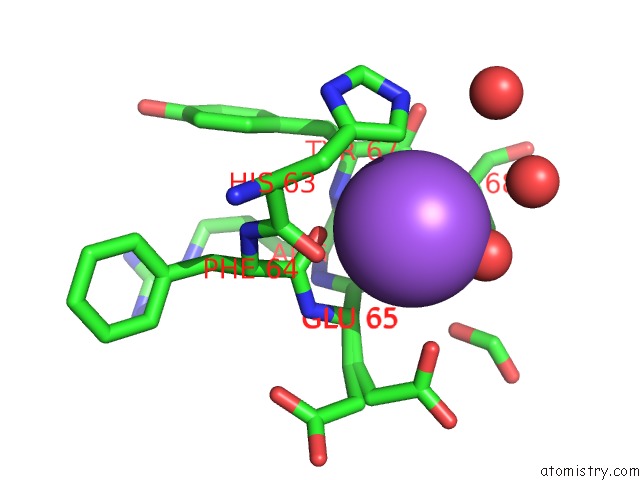
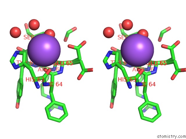
Sodium binding site 1 out of 3 in 5dnu
Go back to
Sodium binding site 1 out
of 3 in the Crystal Structure of Striga KAI2-Like Protein in Complex with Karrikin
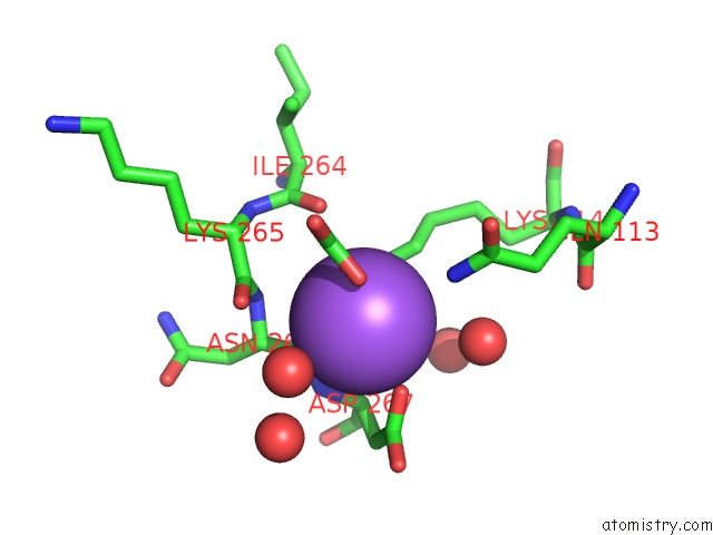
Mono view
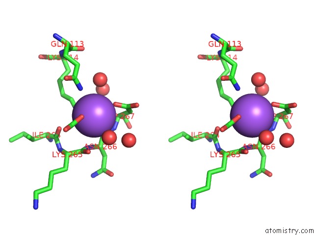
Stereo pair view
Mono view
Stereo pair view
A full contact list of Sodium with other atoms in the Na binding
site number 1 of Crystal Structure of Striga KAI2-Like Protein in Complex with Karrikin within 5.0Å range:
|
Sodium binding site 2 out of 3 in 5dnu
Go back to
Sodium binding site 2 out
of 3 in the Crystal Structure of Striga KAI2-Like Protein in Complex with Karrikin
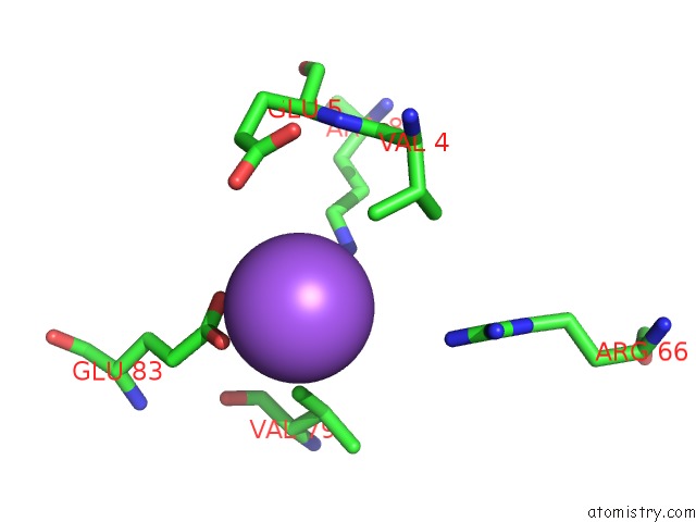
Mono view
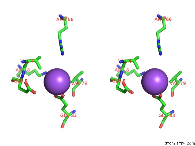
Stereo pair view
Mono view
Stereo pair view
A full contact list of Sodium with other atoms in the Na binding
site number 2 of Crystal Structure of Striga KAI2-Like Protein in Complex with Karrikin within 5.0Å range:
|
Sodium binding site 3 out of 3 in 5dnu
Go back to
Sodium binding site 3 out
of 3 in the Crystal Structure of Striga KAI2-Like Protein in Complex with Karrikin
Mono view
Stereo pair view
Mono view
Stereo pair view
A full contact list of Sodium with other atoms in the Na binding
site number 3 of Crystal Structure of Striga KAI2-Like Protein in Complex with Karrikin within 5.0Å range:
|
Reference:
Y.Xu,
T.Miyakawa,
H.Nakamura,
A.Nakamura,
Y.Imamura,
T.Asami,
M.Tanokura.
Structural Basis of Unique Ligand Specificity of KAI2-Like Protein From Parasitic Weed Striga Hermonthica Sci Rep V. 6 31386 2016.
ISSN: ESSN 2045-2322
PubMed: 27507097
DOI: 10.1038/SREP31386
Page generated: Mon Oct 7 20:39:10 2024
ISSN: ESSN 2045-2322
PubMed: 27507097
DOI: 10.1038/SREP31386
Last articles
Mg in 1VQPMg in 1VQO
Mg in 1W55
Mg in 1W54
Mg in 1W4B
Mg in 1W49
Mg in 1W46
Mg in 1W2Y
Mg in 1VQN
Mg in 1W25