Sodium »
PDB 5ddq-5dy9 »
5dgw »
Sodium in PDB 5dgw: Crystal Structure of Hiv-1 Protease Inhibitor Grl-105-11A Containing Substituted Fused-Tetrahydropyranyl Tetrahydrofuran As P2-Ligand
Protein crystallography data
The structure of Crystal Structure of Hiv-1 Protease Inhibitor Grl-105-11A Containing Substituted Fused-Tetrahydropyranyl Tetrahydrofuran As P2-Ligand, PDB code: 5dgw
was solved by
J.Agniswamy,
Y.-F.Wang,
I.T.Weber,
with X-Ray Crystallography technique. A brief refinement statistics is given in the table below:
| Resolution Low / High (Å) | 50.00 / 1.62 |
| Space group | P 21 21 2 |
| Cell size a, b, c (Å), α, β, γ (°) | 58.677, 86.055, 46.003, 90.00, 90.00, 90.00 |
| R / Rfree (%) | 20.1 / 24.5 |
Other elements in 5dgw:
The structure of Crystal Structure of Hiv-1 Protease Inhibitor Grl-105-11A Containing Substituted Fused-Tetrahydropyranyl Tetrahydrofuran As P2-Ligand also contains other interesting chemical elements:
| Chlorine | (Cl) | 3 atoms |
Sodium Binding Sites:
The binding sites of Sodium atom in the Crystal Structure of Hiv-1 Protease Inhibitor Grl-105-11A Containing Substituted Fused-Tetrahydropyranyl Tetrahydrofuran As P2-Ligand
(pdb code 5dgw). This binding sites where shown within
5.0 Angstroms radius around Sodium atom.
In total only one binding site of Sodium was determined in the Crystal Structure of Hiv-1 Protease Inhibitor Grl-105-11A Containing Substituted Fused-Tetrahydropyranyl Tetrahydrofuran As P2-Ligand, PDB code: 5dgw:
In total only one binding site of Sodium was determined in the Crystal Structure of Hiv-1 Protease Inhibitor Grl-105-11A Containing Substituted Fused-Tetrahydropyranyl Tetrahydrofuran As P2-Ligand, PDB code: 5dgw:
Sodium binding site 1 out of 1 in 5dgw
Go back to
Sodium binding site 1 out
of 1 in the Crystal Structure of Hiv-1 Protease Inhibitor Grl-105-11A Containing Substituted Fused-Tetrahydropyranyl Tetrahydrofuran As P2-Ligand
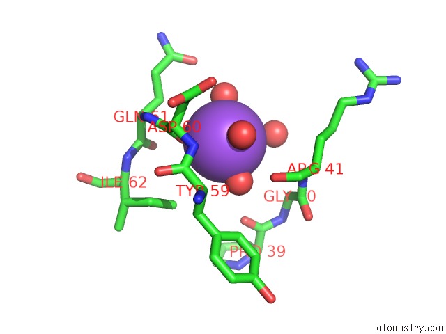
Mono view
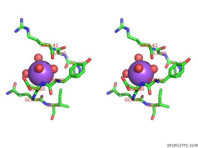
Stereo pair view
Mono view
Stereo pair view
A full contact list of Sodium with other atoms in the Na binding
site number 1 of Crystal Structure of Hiv-1 Protease Inhibitor Grl-105-11A Containing Substituted Fused-Tetrahydropyranyl Tetrahydrofuran As P2-Ligand within 5.0Å range:
|
Reference:
A.K.Ghosh,
C.D.Martyr,
L.A.Kassekert,
P.R.Nyalapatla,
M.Steffey,
J.Agniswamy,
Y.F.Wang,
I.T.Weber,
M.Amano,
H.Mitsuya.
Design, Synthesis, Biological Evaluation and X-Ray Structural Studies of Hiv-1 Protease Inhibitors Containing Substituted Fused-Tetrahydropyranyl Tetrahydrofuran As P2-Ligands. Org.Biomol.Chem. V. 13 11607 2015.
ISSN: ESSN 1477-0539
PubMed: 26462551
DOI: 10.1039/C5OB01930C
Page generated: Mon Oct 7 20:36:24 2024
ISSN: ESSN 1477-0539
PubMed: 26462551
DOI: 10.1039/C5OB01930C
Last articles
Mg in 4FVUMg in 4FWI
Mg in 4FVQ
Mg in 4FVR
Mg in 4FUT
Mg in 4FS5
Mg in 4FRG
Mg in 4FS2
Mg in 4FMA
Mg in 4FR8