Sodium »
PDB 4yil-4z48 »
4yt9 »
Sodium in PDB 4yt9: Crystal Structure of Porphyromonas Gingivalis Peptidylarginine Deiminase (Ppad) Substrate-Unbound.
Protein crystallography data
The structure of Crystal Structure of Porphyromonas Gingivalis Peptidylarginine Deiminase (Ppad) Substrate-Unbound., PDB code: 4yt9
was solved by
T.Goulas,
D.Mizgalska,
I.Garcia-Ferrer,
T.Kantyka,
T.Guevara,
B.Szmigielski,
A.Sroka,
C.Millan,
I.Uson,
F.Veillard,
B.Potempa,
P.Mydel,
M.Sola,
J.Potempa,
F.X.Gomis-Ruth,
with X-Ray Crystallography technique. A brief refinement statistics is given in the table below:
| Resolution Low / High (Å) | 42.01 / 1.50 |
| Space group | P 21 21 21 |
| Cell size a, b, c (Å), α, β, γ (°) | 58.560, 60.300, 113.680, 90.00, 90.00, 90.00 |
| R / Rfree (%) | 15.7 / 17.7 |
Sodium Binding Sites:
The binding sites of Sodium atom in the Crystal Structure of Porphyromonas Gingivalis Peptidylarginine Deiminase (Ppad) Substrate-Unbound.
(pdb code 4yt9). This binding sites where shown within
5.0 Angstroms radius around Sodium atom.
In total only one binding site of Sodium was determined in the Crystal Structure of Porphyromonas Gingivalis Peptidylarginine Deiminase (Ppad) Substrate-Unbound., PDB code: 4yt9:
In total only one binding site of Sodium was determined in the Crystal Structure of Porphyromonas Gingivalis Peptidylarginine Deiminase (Ppad) Substrate-Unbound., PDB code: 4yt9:
Sodium binding site 1 out of 1 in 4yt9
Go back to
Sodium binding site 1 out
of 1 in the Crystal Structure of Porphyromonas Gingivalis Peptidylarginine Deiminase (Ppad) Substrate-Unbound.
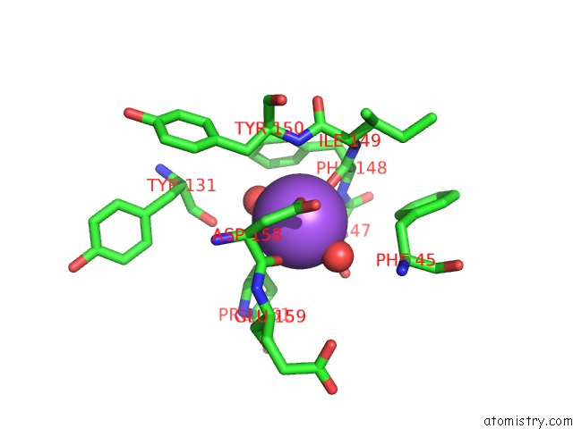
Mono view
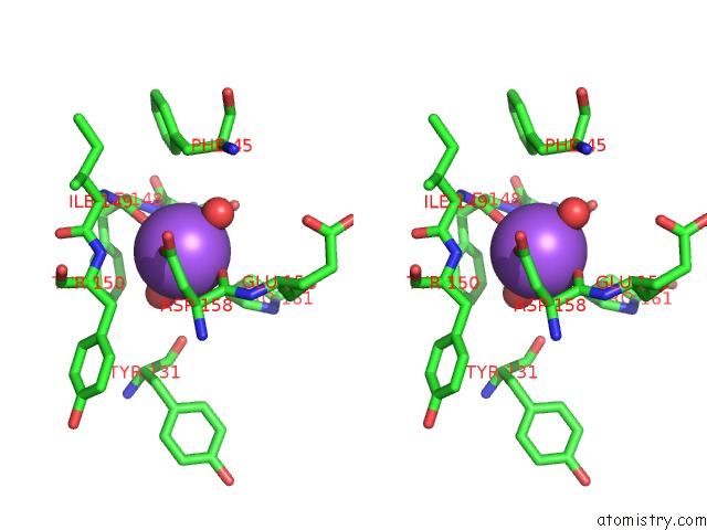
Stereo pair view
Mono view
Stereo pair view
A full contact list of Sodium with other atoms in the Na binding
site number 1 of Crystal Structure of Porphyromonas Gingivalis Peptidylarginine Deiminase (Ppad) Substrate-Unbound. within 5.0Å range:
|
Reference:
T.Goulas,
D.Mizgalska,
I.Garcia-Ferrer,
T.Kantyka,
T.Guevara,
B.Szmigielski,
A.Sroka,
C.Millan,
I.Uson,
F.Veillard,
B.Potempa,
P.Mydel,
M.Sola,
J.Potempa,
F.X.Gomis-Ruth.
Structure and Mechanism of A Bacterial Host-Protein Citrullinating Virulence Factor, Porphyromonas Gingivalis Peptidylarginine Deiminase. Sci Rep V. 5 11969 2015.
ISSN: ESSN 2045-2322
PubMed: 26132828
DOI: 10.1038/SREP11969
Page generated: Sun Aug 17 22:39:45 2025
ISSN: ESSN 2045-2322
PubMed: 26132828
DOI: 10.1038/SREP11969
Last articles
K in 9NESK in 9PHG
K in 9NEI
K in 9NED
K in 9NEC
K in 9NEG
K in 9CWU
K in 9CVB
K in 9CVA
K in 9COM