Sodium »
PDB 4rne-4tmv »
4tkx »
Sodium in PDB 4tkx: Structure of Protease
Enzymatic activity of Structure of Protease
All present enzymatic activity of Structure of Protease:
3.4.22.47;
3.4.22.47;
Protein crystallography data
The structure of Structure of Protease, PDB code: 4tkx
was solved by
M.A.Gorman,
M.W.Parker,
with X-Ray Crystallography technique. A brief refinement statistics is given in the table below:
| Resolution Low / High (Å) | 40.71 / 1.60 |
| Space group | P 43 21 2 |
| Cell size a, b, c (Å), α, β, γ (°) | 116.941, 116.941, 86.852, 90.00, 90.00, 90.00 |
| R / Rfree (%) | 12 / 14.5 |
Other elements in 4tkx:
The structure of Structure of Protease also contains other interesting chemical elements:
| Lead | (Pb) | 1 atom |
| Potassium | (K) | 1 atom |
Sodium Binding Sites:
The binding sites of Sodium atom in the Structure of Protease
(pdb code 4tkx). This binding sites where shown within
5.0 Angstroms radius around Sodium atom.
In total 2 binding sites of Sodium where determined in the Structure of Protease, PDB code: 4tkx:
Jump to Sodium binding site number: 1; 2;
In total 2 binding sites of Sodium where determined in the Structure of Protease, PDB code: 4tkx:
Jump to Sodium binding site number: 1; 2;
Sodium binding site 1 out of 2 in 4tkx
Go back to
Sodium binding site 1 out
of 2 in the Structure of Protease
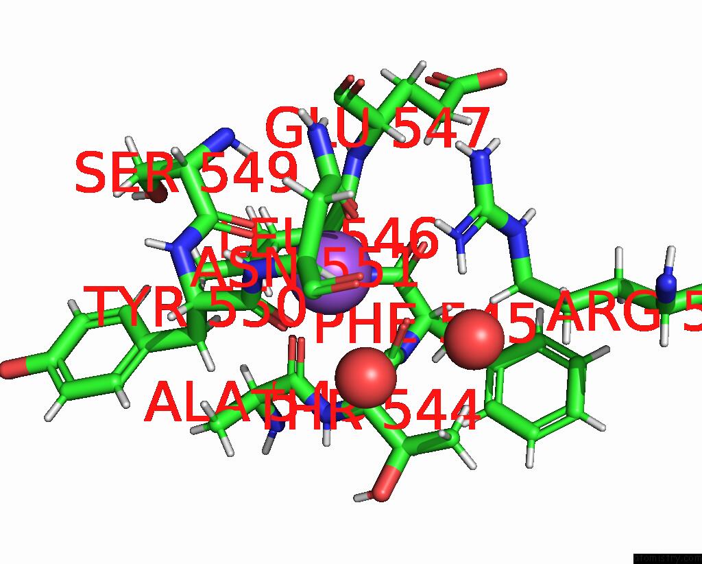
Mono view
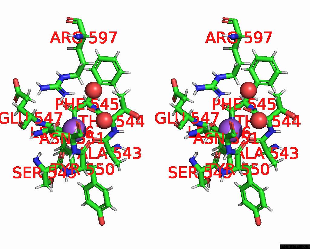
Stereo pair view
Mono view
Stereo pair view
A full contact list of Sodium with other atoms in the Na binding
site number 1 of Structure of Protease within 5.0Å range:
|
Sodium binding site 2 out of 2 in 4tkx
Go back to
Sodium binding site 2 out
of 2 in the Structure of Protease
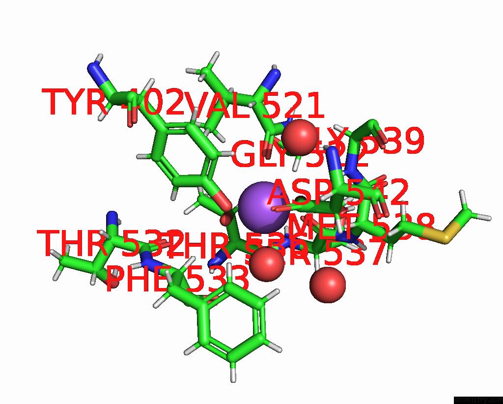
Mono view
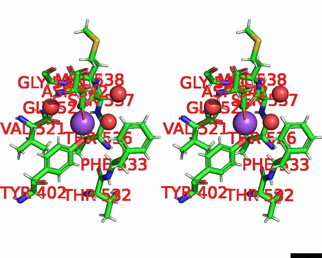
Stereo pair view
Mono view
Stereo pair view
A full contact list of Sodium with other atoms in the Na binding
site number 2 of Structure of Protease within 5.0Å range:
|
Reference:
M.A.Gorman,
C.A.Seers,
B.J.Michell,
S.C.Feil,
N.L.Huq,
K.J.Cross,
E.C.Reynolds,
M.W.Parker.
Structure of the Lysine Specific Protease Kgp From Porphyromonas Gingivalis, A Target For Improved Oral Health. Protein Sci. V. 24 162 2015.
ISSN: ESSN 1469-896X
PubMed: 25327141
DOI: 10.1002/PRO.2589
Page generated: Mon Oct 7 18:24:39 2024
ISSN: ESSN 1469-896X
PubMed: 25327141
DOI: 10.1002/PRO.2589
Last articles
Mg in 5ZKJMg in 5ZKI
Mg in 5ZK6
Mg in 5ZE9
Mg in 5ZFX
Mg in 5ZCT
Mg in 5ZE6
Mg in 5ZE4
Mg in 5ZDN
Mg in 5ZE0