Sodium »
PDB 4pd8-4pp4 »
4pf4 »
Sodium in PDB 4pf4: 1.1A X-Ray Structure of the Apo Catalytic Domain of Death-Associated Protein Kinase 1, Aa 1-277
Enzymatic activity of 1.1A X-Ray Structure of the Apo Catalytic Domain of Death-Associated Protein Kinase 1, Aa 1-277
All present enzymatic activity of 1.1A X-Ray Structure of the Apo Catalytic Domain of Death-Associated Protein Kinase 1, Aa 1-277:
2.7.11.1;
2.7.11.1;
Protein crystallography data
The structure of 1.1A X-Ray Structure of the Apo Catalytic Domain of Death-Associated Protein Kinase 1, Aa 1-277, PDB code: 4pf4
was solved by
K.Temmerman,
B.Simon,
M.Wilmanns,
with X-Ray Crystallography technique. A brief refinement statistics is given in the table below:
| Resolution Low / High (Å) | 50.76 / 1.13 |
| Space group | P 21 21 21 |
| Cell size a, b, c (Å), α, β, γ (°) | 46.516, 62.005, 88.375, 90.00, 90.00, 90.00 |
| R / Rfree (%) | 11.7 / 15 |
Other elements in 4pf4:
The structure of 1.1A X-Ray Structure of the Apo Catalytic Domain of Death-Associated Protein Kinase 1, Aa 1-277 also contains other interesting chemical elements:
| Magnesium | (Mg) | 1 atom |
Sodium Binding Sites:
The binding sites of Sodium atom in the 1.1A X-Ray Structure of the Apo Catalytic Domain of Death-Associated Protein Kinase 1, Aa 1-277
(pdb code 4pf4). This binding sites where shown within
5.0 Angstroms radius around Sodium atom.
In total 2 binding sites of Sodium where determined in the 1.1A X-Ray Structure of the Apo Catalytic Domain of Death-Associated Protein Kinase 1, Aa 1-277, PDB code: 4pf4:
Jump to Sodium binding site number: 1; 2;
In total 2 binding sites of Sodium where determined in the 1.1A X-Ray Structure of the Apo Catalytic Domain of Death-Associated Protein Kinase 1, Aa 1-277, PDB code: 4pf4:
Jump to Sodium binding site number: 1; 2;
Sodium binding site 1 out of 2 in 4pf4
Go back to
Sodium binding site 1 out
of 2 in the 1.1A X-Ray Structure of the Apo Catalytic Domain of Death-Associated Protein Kinase 1, Aa 1-277
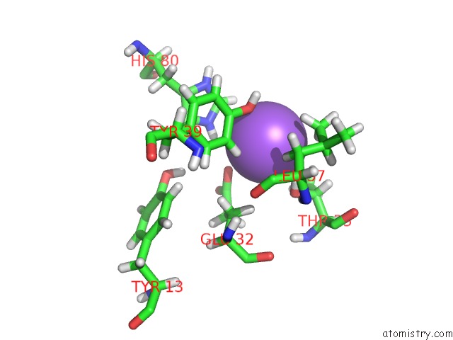
Mono view
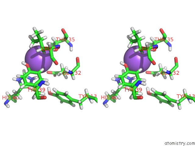
Stereo pair view
Mono view
Stereo pair view
A full contact list of Sodium with other atoms in the Na binding
site number 1 of 1.1A X-Ray Structure of the Apo Catalytic Domain of Death-Associated Protein Kinase 1, Aa 1-277 within 5.0Å range:
|
Sodium binding site 2 out of 2 in 4pf4
Go back to
Sodium binding site 2 out
of 2 in the 1.1A X-Ray Structure of the Apo Catalytic Domain of Death-Associated Protein Kinase 1, Aa 1-277
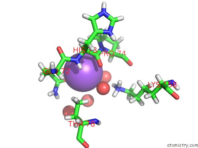
Mono view
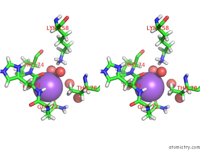
Stereo pair view
Mono view
Stereo pair view
A full contact list of Sodium with other atoms in the Na binding
site number 2 of 1.1A X-Ray Structure of the Apo Catalytic Domain of Death-Associated Protein Kinase 1, Aa 1-277 within 5.0Å range:
|
Reference:
K.Temmerman,
B.Simon,
M.Wilmanns.
1.1A X-Ray Structure of the Apo Catalytic Domain of Death-Associated Protein Kinase 1, Aa 1-277 To Be Published.
Page generated: Mon Oct 7 17:42:09 2024
Last articles
Mg in 5EKEMg in 5EJZ
Mg in 5EIY
Mg in 5EJ0
Mg in 5EHH
Mg in 5EIB
Mg in 5EGY
Mg in 5EIK
Mg in 5E7C
Mg in 5EFQ