Sodium »
PDB 4oci-4owc »
4oub »
Sodium in PDB 4oub: A 2.20 Angstroms X-Ray Crystal Structure of E268A 2-Aminomucaonate 6- Semialdehyde Dehydrogenase Catalytic Intermediate From Pseudomonas Fluorescens
Protein crystallography data
The structure of A 2.20 Angstroms X-Ray Crystal Structure of E268A 2-Aminomucaonate 6- Semialdehyde Dehydrogenase Catalytic Intermediate From Pseudomonas Fluorescens, PDB code: 4oub
was solved by
L.Huo,
A.Liu,
with X-Ray Crystallography technique. A brief refinement statistics is given in the table below:
| Resolution Low / High (Å) | 41.00 / 2.19 |
| Space group | P 21 21 21 |
| Cell size a, b, c (Å), α, β, γ (°) | 88.329, 141.345, 173.530, 90.00, 90.00, 90.00 |
| R / Rfree (%) | 18 / 22.5 |
Sodium Binding Sites:
The binding sites of Sodium atom in the A 2.20 Angstroms X-Ray Crystal Structure of E268A 2-Aminomucaonate 6- Semialdehyde Dehydrogenase Catalytic Intermediate From Pseudomonas Fluorescens
(pdb code 4oub). This binding sites where shown within
5.0 Angstroms radius around Sodium atom.
In total 4 binding sites of Sodium where determined in the A 2.20 Angstroms X-Ray Crystal Structure of E268A 2-Aminomucaonate 6- Semialdehyde Dehydrogenase Catalytic Intermediate From Pseudomonas Fluorescens, PDB code: 4oub:
Jump to Sodium binding site number: 1; 2; 3; 4;
In total 4 binding sites of Sodium where determined in the A 2.20 Angstroms X-Ray Crystal Structure of E268A 2-Aminomucaonate 6- Semialdehyde Dehydrogenase Catalytic Intermediate From Pseudomonas Fluorescens, PDB code: 4oub:
Jump to Sodium binding site number: 1; 2; 3; 4;
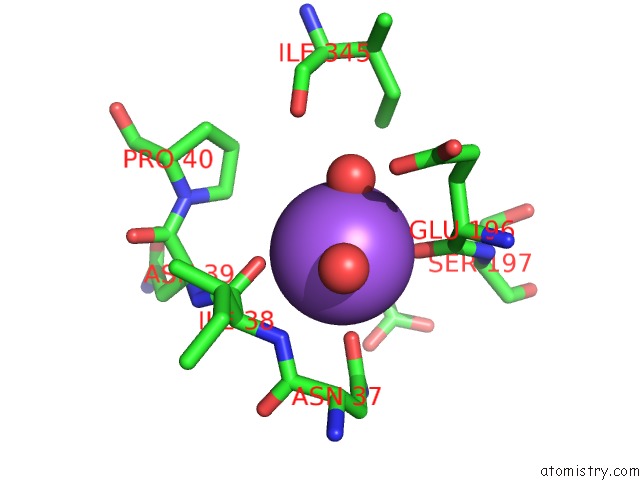
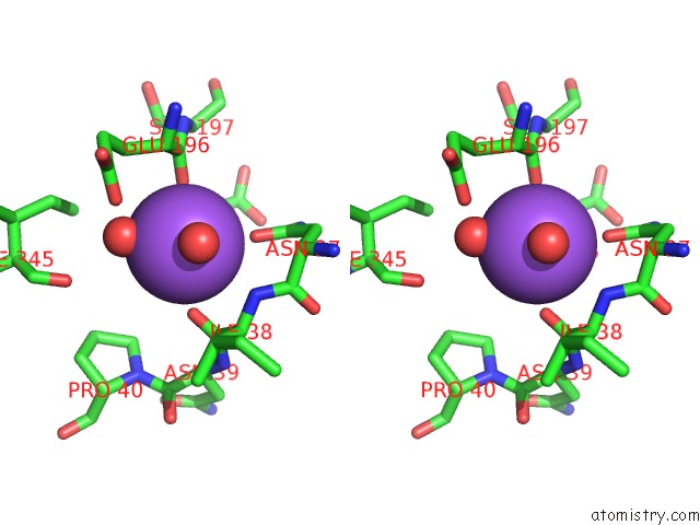
Sodium binding site 1 out of 4 in 4oub
Go back to
Sodium binding site 1 out
of 4 in the A 2.20 Angstroms X-Ray Crystal Structure of E268A 2-Aminomucaonate 6- Semialdehyde Dehydrogenase Catalytic Intermediate From Pseudomonas Fluorescens
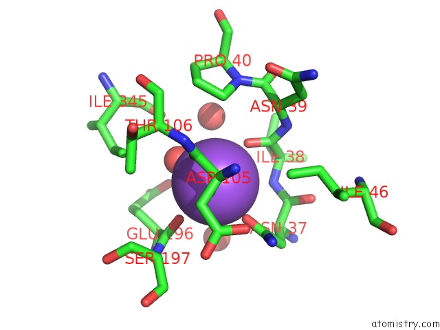
Mono view
Stereo pair view
Mono view
Stereo pair view
A full contact list of Sodium with other atoms in the Na binding
site number 1 of A 2.20 Angstroms X-Ray Crystal Structure of E268A 2-Aminomucaonate 6- Semialdehyde Dehydrogenase Catalytic Intermediate From Pseudomonas Fluorescens within 5.0Å range:
|
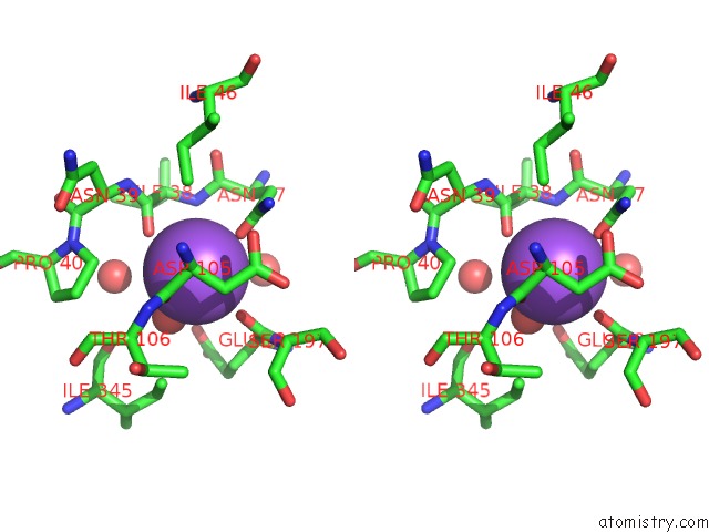
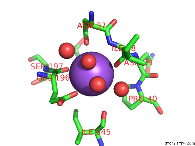
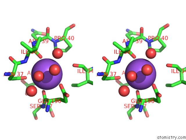
Sodium binding site 2 out of 4 in 4oub
Go back to
Sodium binding site 2 out
of 4 in the A 2.20 Angstroms X-Ray Crystal Structure of E268A 2-Aminomucaonate 6- Semialdehyde Dehydrogenase Catalytic Intermediate From Pseudomonas Fluorescens
Mono view
Stereo pair view
Mono view
Stereo pair view
A full contact list of Sodium with other atoms in the Na binding
site number 2 of A 2.20 Angstroms X-Ray Crystal Structure of E268A 2-Aminomucaonate 6- Semialdehyde Dehydrogenase Catalytic Intermediate From Pseudomonas Fluorescens within 5.0Å range:
|
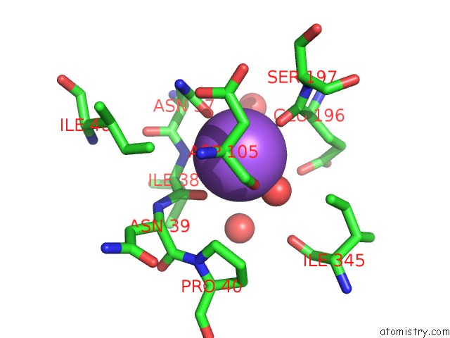
Sodium binding site 3 out of 4 in 4oub
Go back to
Sodium binding site 3 out
of 4 in the A 2.20 Angstroms X-Ray Crystal Structure of E268A 2-Aminomucaonate 6- Semialdehyde Dehydrogenase Catalytic Intermediate From Pseudomonas Fluorescens
Mono view
Stereo pair view
Mono view
Stereo pair view
A full contact list of Sodium with other atoms in the Na binding
site number 3 of A 2.20 Angstroms X-Ray Crystal Structure of E268A 2-Aminomucaonate 6- Semialdehyde Dehydrogenase Catalytic Intermediate From Pseudomonas Fluorescens within 5.0Å range:
|
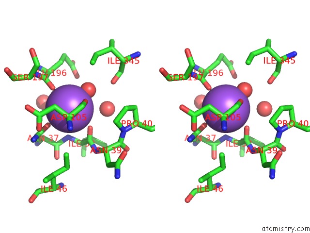
Sodium binding site 4 out of 4 in 4oub
Go back to
Sodium binding site 4 out
of 4 in the A 2.20 Angstroms X-Ray Crystal Structure of E268A 2-Aminomucaonate 6- Semialdehyde Dehydrogenase Catalytic Intermediate From Pseudomonas Fluorescens
Mono view
Stereo pair view
Mono view
Stereo pair view
A full contact list of Sodium with other atoms in the Na binding
site number 4 of A 2.20 Angstroms X-Ray Crystal Structure of E268A 2-Aminomucaonate 6- Semialdehyde Dehydrogenase Catalytic Intermediate From Pseudomonas Fluorescens within 5.0Å range:
|
Reference:
L.Huo,
I.Davis,
F.Liu,
H.Iwaki,
Y.Hasegawa,
A.Liu.
Crystallographic and Spectroscopic Snapshots Reveal A Dehydrogenase in Action Nat Commun 2014.
ISSN: ESSN 2041-1723
DOI: 10.1038/NCOMMS6935
Page generated: Mon Oct 7 17:33:25 2024
ISSN: ESSN 2041-1723
DOI: 10.1038/NCOMMS6935
Last articles
Mg in 4AS1Mg in 4ARZ
Mg in 4ARQ
Mg in 4ARK
Mg in 4ARI
Mg in 4AQX
Mg in 4ARC
Mg in 4AQV
Mg in 4AQW
Mg in 4AQ7