Sodium »
PDB 4mfc-4mzx »
4mr1 »
Sodium in PDB 4mr1: X-Ray Structure of the Adduct Between Hen Egg White Lysozyme and Cis- Diamminediiodoplatinum(II)
Enzymatic activity of X-Ray Structure of the Adduct Between Hen Egg White Lysozyme and Cis- Diamminediiodoplatinum(II)
All present enzymatic activity of X-Ray Structure of the Adduct Between Hen Egg White Lysozyme and Cis- Diamminediiodoplatinum(II):
3.2.1.17;
3.2.1.17;
Protein crystallography data
The structure of X-Ray Structure of the Adduct Between Hen Egg White Lysozyme and Cis- Diamminediiodoplatinum(II), PDB code: 4mr1
was solved by
A.Merlino,
with X-Ray Crystallography technique. A brief refinement statistics is given in the table below:
| Resolution Low / High (Å) | 27.23 / 1.99 |
| Space group | P 43 21 2 |
| Cell size a, b, c (Å), α, β, γ (°) | 77.017, 77.017, 36.834, 90.00, 90.00, 90.00 |
| R / Rfree (%) | 18.4 / 23.4 |
Other elements in 4mr1:
The structure of X-Ray Structure of the Adduct Between Hen Egg White Lysozyme and Cis- Diamminediiodoplatinum(II) also contains other interesting chemical elements:
| Platinum | (Pt) | 2 atoms |
| Iodine | (I) | 4 atoms |
| Chlorine | (Cl) | 2 atoms |
Sodium Binding Sites:
The binding sites of Sodium atom in the X-Ray Structure of the Adduct Between Hen Egg White Lysozyme and Cis- Diamminediiodoplatinum(II)
(pdb code 4mr1). This binding sites where shown within
5.0 Angstroms radius around Sodium atom.
In total only one binding site of Sodium was determined in the X-Ray Structure of the Adduct Between Hen Egg White Lysozyme and Cis- Diamminediiodoplatinum(II), PDB code: 4mr1:
In total only one binding site of Sodium was determined in the X-Ray Structure of the Adduct Between Hen Egg White Lysozyme and Cis- Diamminediiodoplatinum(II), PDB code: 4mr1:
Sodium binding site 1 out of 1 in 4mr1
Go back to
Sodium binding site 1 out
of 1 in the X-Ray Structure of the Adduct Between Hen Egg White Lysozyme and Cis- Diamminediiodoplatinum(II)

Mono view
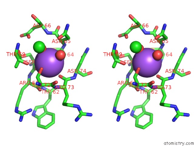
Stereo pair view
Mono view
Stereo pair view
A full contact list of Sodium with other atoms in the Na binding
site number 1 of X-Ray Structure of the Adduct Between Hen Egg White Lysozyme and Cis- Diamminediiodoplatinum(II) within 5.0Å range:
|
Reference:
L.Messori,
T.Marzo,
C.Gabbiani,
A.A.Valdes,
A.G.Quiroga,
A.Merlino.
Peculiar Features in the Crystal Structure of the Adduct Formed Between Cis-PTI2(NH3)2 and Hen Egg White Lysozyme. Inorg.Chem. V. 52 13827 2013.
ISSN: ISSN 0020-1669
PubMed: 24256441
DOI: 10.1021/IC402611M
Page generated: Sun Aug 17 20:40:44 2025
ISSN: ISSN 0020-1669
PubMed: 24256441
DOI: 10.1021/IC402611M
Last articles
Mn in 9LJUMn in 9LJW
Mn in 9LJS
Mn in 9LJR
Mn in 9LJT
Mn in 9LJV
Mg in 9UA2
Mg in 9R96
Mg in 9VM1
Mg in 9P01