Sodium »
PDB 4kxy-4lgn »
4l1q »
Sodium in PDB 4l1q: Crystal Structure of the E113Q-Maug/Pre-Methylamine Dehydrogenase Complex
Enzymatic activity of Crystal Structure of the E113Q-Maug/Pre-Methylamine Dehydrogenase Complex
All present enzymatic activity of Crystal Structure of the E113Q-Maug/Pre-Methylamine Dehydrogenase Complex:
1.4.99.3;
1.4.99.3;
Protein crystallography data
The structure of Crystal Structure of the E113Q-Maug/Pre-Methylamine Dehydrogenase Complex, PDB code: 4l1q
was solved by
E.Y.Yukl,
C.M.Wilmot,
with X-Ray Crystallography technique. A brief refinement statistics is given in the table below:
| Resolution Low / High (Å) | 24.35 / 1.92 |
| Space group | P 1 |
| Cell size a, b, c (Å), α, β, γ (°) | 55.530, 83.520, 107.780, 109.94, 91.54, 105.78 |
| R / Rfree (%) | 16 / 20.7 |
Other elements in 4l1q:
The structure of Crystal Structure of the E113Q-Maug/Pre-Methylamine Dehydrogenase Complex also contains other interesting chemical elements:
| Iron | (Fe) | 4 atoms |
| Calcium | (Ca) | 2 atoms |
Sodium Binding Sites:
The binding sites of Sodium atom in the Crystal Structure of the E113Q-Maug/Pre-Methylamine Dehydrogenase Complex
(pdb code 4l1q). This binding sites where shown within
5.0 Angstroms radius around Sodium atom.
In total 3 binding sites of Sodium where determined in the Crystal Structure of the E113Q-Maug/Pre-Methylamine Dehydrogenase Complex, PDB code: 4l1q:
Jump to Sodium binding site number: 1; 2; 3;
In total 3 binding sites of Sodium where determined in the Crystal Structure of the E113Q-Maug/Pre-Methylamine Dehydrogenase Complex, PDB code: 4l1q:
Jump to Sodium binding site number: 1; 2; 3;
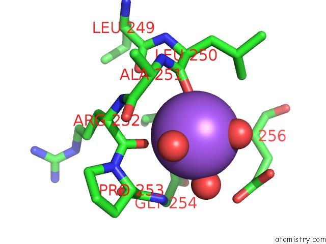
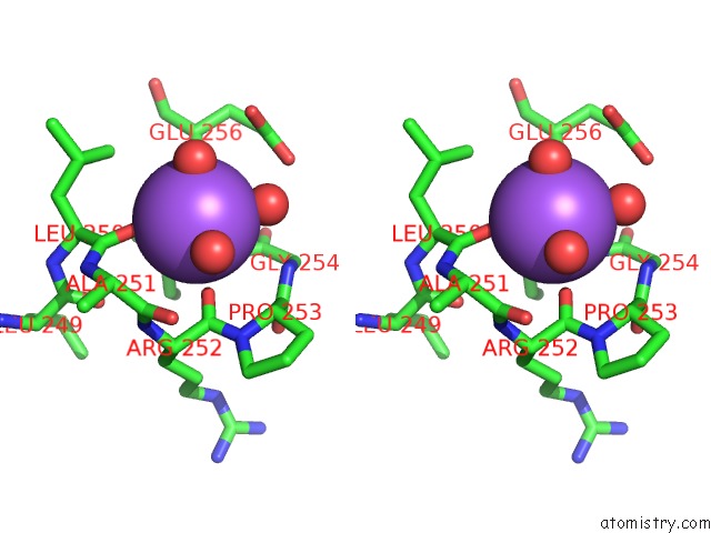
Sodium binding site 1 out of 3 in 4l1q
Go back to
Sodium binding site 1 out
of 3 in the Crystal Structure of the E113Q-Maug/Pre-Methylamine Dehydrogenase Complex
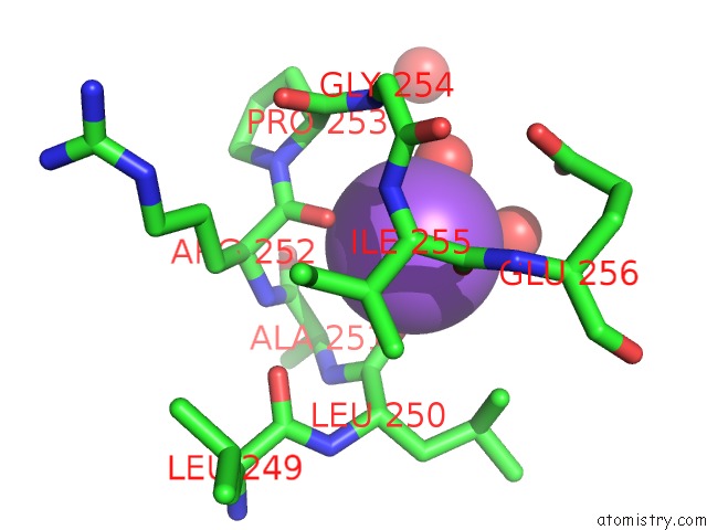
Mono view
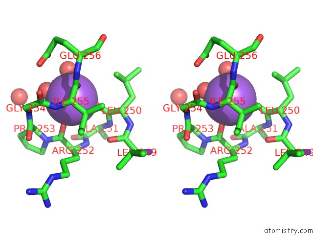
Stereo pair view
Mono view
Stereo pair view
A full contact list of Sodium with other atoms in the Na binding
site number 1 of Crystal Structure of the E113Q-Maug/Pre-Methylamine Dehydrogenase Complex within 5.0Å range:
|
Sodium binding site 2 out of 3 in 4l1q
Go back to
Sodium binding site 2 out
of 3 in the Crystal Structure of the E113Q-Maug/Pre-Methylamine Dehydrogenase Complex
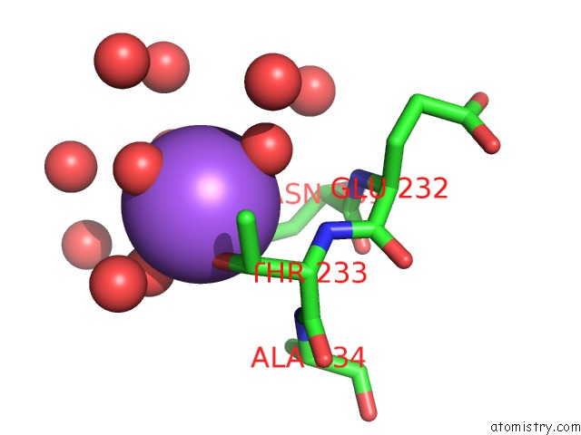
Mono view
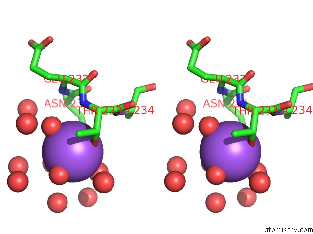
Stereo pair view
Mono view
Stereo pair view
A full contact list of Sodium with other atoms in the Na binding
site number 2 of Crystal Structure of the E113Q-Maug/Pre-Methylamine Dehydrogenase Complex within 5.0Å range:
|
Sodium binding site 3 out of 3 in 4l1q
Go back to
Sodium binding site 3 out
of 3 in the Crystal Structure of the E113Q-Maug/Pre-Methylamine Dehydrogenase Complex
Mono view
Stereo pair view
Mono view
Stereo pair view
A full contact list of Sodium with other atoms in the Na binding
site number 3 of Crystal Structure of the E113Q-Maug/Pre-Methylamine Dehydrogenase Complex within 5.0Å range:
|
Reference:
N.Abu Tarboush,
E.T.Yukl,
S.Shin,
M.Feng,
C.M.Wilmot,
V.L.Davidson.
Carboxyl Group of GLU113 Is Required For Stabilization of the Diferrous and Bis-Fe(IV) States of Maug. Biochemistry V. 52 6358 2013.
ISSN: ISSN 0006-2960
PubMed: 23952537
DOI: 10.1021/BI400905S
Page generated: Mon Oct 7 16:40:16 2024
ISSN: ISSN 0006-2960
PubMed: 23952537
DOI: 10.1021/BI400905S
Last articles
I in 3WN5I in 3WYX
I in 3WGW
I in 3WD6
I in 3WB5
I in 3W31
I in 3WB4
I in 3W1N
I in 3W0F
I in 3W2Z