Sodium »
PDB 4i0w-4iib »
4i25 »
Sodium in PDB 4i25: 2.00 Angstroms X-Ray Crystal Structure of Nad- and Substrate-Bound 2- Aminomuconate 6-Semialdehyde Dehydrogenase From Pseudomonas Fluorescens
Protein crystallography data
The structure of 2.00 Angstroms X-Ray Crystal Structure of Nad- and Substrate-Bound 2- Aminomuconate 6-Semialdehyde Dehydrogenase From Pseudomonas Fluorescens, PDB code: 4i25
was solved by
L.Huo,
I.Davis,
L.Chen,
A.Liu,
with X-Ray Crystallography technique. A brief refinement statistics is given in the table below:
| Resolution Low / High (Å) | 27.22 / 2.00 |
| Space group | P 21 21 21 |
| Cell size a, b, c (Å), α, β, γ (°) | 88.253, 142.932, 175.125, 90.00, 90.00, 90.00 |
| R / Rfree (%) | 16.2 / 20.8 |
Sodium Binding Sites:
The binding sites of Sodium atom in the 2.00 Angstroms X-Ray Crystal Structure of Nad- and Substrate-Bound 2- Aminomuconate 6-Semialdehyde Dehydrogenase From Pseudomonas Fluorescens
(pdb code 4i25). This binding sites where shown within
5.0 Angstroms radius around Sodium atom.
In total 4 binding sites of Sodium where determined in the 2.00 Angstroms X-Ray Crystal Structure of Nad- and Substrate-Bound 2- Aminomuconate 6-Semialdehyde Dehydrogenase From Pseudomonas Fluorescens, PDB code: 4i25:
Jump to Sodium binding site number: 1; 2; 3; 4;
In total 4 binding sites of Sodium where determined in the 2.00 Angstroms X-Ray Crystal Structure of Nad- and Substrate-Bound 2- Aminomuconate 6-Semialdehyde Dehydrogenase From Pseudomonas Fluorescens, PDB code: 4i25:
Jump to Sodium binding site number: 1; 2; 3; 4;
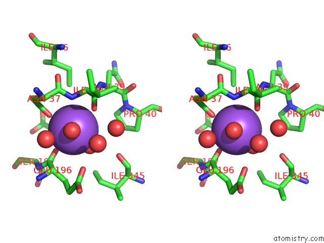
Sodium binding site 1 out of 4 in 4i25
Go back to
Sodium binding site 1 out
of 4 in the 2.00 Angstroms X-Ray Crystal Structure of Nad- and Substrate-Bound 2- Aminomuconate 6-Semialdehyde Dehydrogenase From Pseudomonas Fluorescens

Mono view

Stereo pair view
Mono view
Stereo pair view
A full contact list of Sodium with other atoms in the Na binding
site number 1 of 2.00 Angstroms X-Ray Crystal Structure of Nad- and Substrate-Bound 2- Aminomuconate 6-Semialdehyde Dehydrogenase From Pseudomonas Fluorescens within 5.0Å range:
|
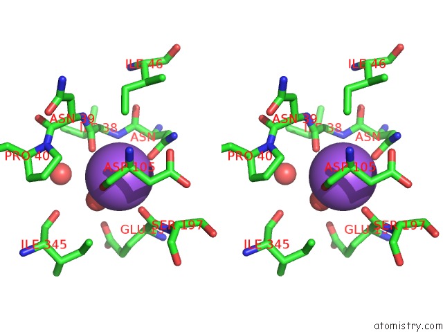
Sodium binding site 2 out of 4 in 4i25
Go back to
Sodium binding site 2 out
of 4 in the 2.00 Angstroms X-Ray Crystal Structure of Nad- and Substrate-Bound 2- Aminomuconate 6-Semialdehyde Dehydrogenase From Pseudomonas Fluorescens

Mono view
Stereo pair view
Mono view
Stereo pair view
A full contact list of Sodium with other atoms in the Na binding
site number 2 of 2.00 Angstroms X-Ray Crystal Structure of Nad- and Substrate-Bound 2- Aminomuconate 6-Semialdehyde Dehydrogenase From Pseudomonas Fluorescens within 5.0Å range:
|
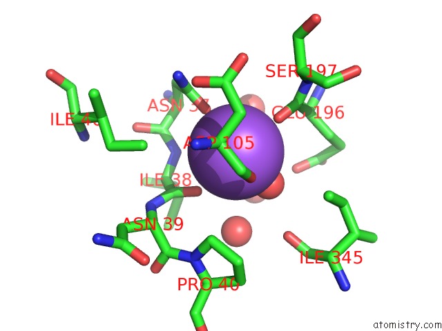
Sodium binding site 3 out of 4 in 4i25
Go back to
Sodium binding site 3 out
of 4 in the 2.00 Angstroms X-Ray Crystal Structure of Nad- and Substrate-Bound 2- Aminomuconate 6-Semialdehyde Dehydrogenase From Pseudomonas Fluorescens

Mono view
Stereo pair view
Mono view
Stereo pair view
A full contact list of Sodium with other atoms in the Na binding
site number 3 of 2.00 Angstroms X-Ray Crystal Structure of Nad- and Substrate-Bound 2- Aminomuconate 6-Semialdehyde Dehydrogenase From Pseudomonas Fluorescens within 5.0Å range:
|
Sodium binding site 4 out of 4 in 4i25
Go back to
Sodium binding site 4 out
of 4 in the 2.00 Angstroms X-Ray Crystal Structure of Nad- and Substrate-Bound 2- Aminomuconate 6-Semialdehyde Dehydrogenase From Pseudomonas Fluorescens
Mono view

Stereo pair view
Mono view
Stereo pair view
A full contact list of Sodium with other atoms in the Na binding
site number 4 of 2.00 Angstroms X-Ray Crystal Structure of Nad- and Substrate-Bound 2- Aminomuconate 6-Semialdehyde Dehydrogenase From Pseudomonas Fluorescens within 5.0Å range:
|
Reference:
L.Huo,
I.Davis,
L.Chen,
A.Liu.
2.00 Angstroms X-Ray Crystal Structure of Nad- and Substrate-Bound 2-Aminomuconate 6-Semialdehyde Dehydrogenase From Pseudomonas Fluorescens To Be Published.
Page generated: Sun Aug 17 19:45:34 2025
Last articles
Mn in 9LJUMn in 9LJW
Mn in 9LJS
Mn in 9LJR
Mn in 9LJT
Mn in 9LJV
Mg in 9UA2
Mg in 9R96
Mg in 9VM1
Mg in 9P01