Sodium »
PDB 4fpa-4gfi »
4fud »
Sodium in PDB 4fud: Crystal Structure of the Urokinase
Enzymatic activity of Crystal Structure of the Urokinase
All present enzymatic activity of Crystal Structure of the Urokinase:
3.4.21.73;
3.4.21.73;
Protein crystallography data
The structure of Crystal Structure of the Urokinase, PDB code: 4fud
was solved by
Y.N.Kang,
J.A.Stuckey,
V.Nienaber,
V.Giranda,
with X-Ray Crystallography technique. A brief refinement statistics is given in the table below:
| Resolution Low / High (Å) | 27.56 / 2.00 |
| Space group | P 21 21 21 |
| Cell size a, b, c (Å), α, β, γ (°) | 55.000, 52.600, 80.000, 90.00, 90.00, 90.00 |
| R / Rfree (%) | 17.1 / 22 |
Sodium Binding Sites:
The binding sites of Sodium atom in the Crystal Structure of the Urokinase
(pdb code 4fud). This binding sites where shown within
5.0 Angstroms radius around Sodium atom.
In total only one binding site of Sodium was determined in the Crystal Structure of the Urokinase, PDB code: 4fud:
In total only one binding site of Sodium was determined in the Crystal Structure of the Urokinase, PDB code: 4fud:
Sodium binding site 1 out of 1 in 4fud
Go back to
Sodium binding site 1 out
of 1 in the Crystal Structure of the Urokinase
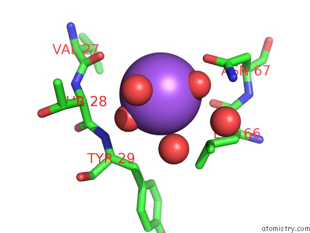
Mono view
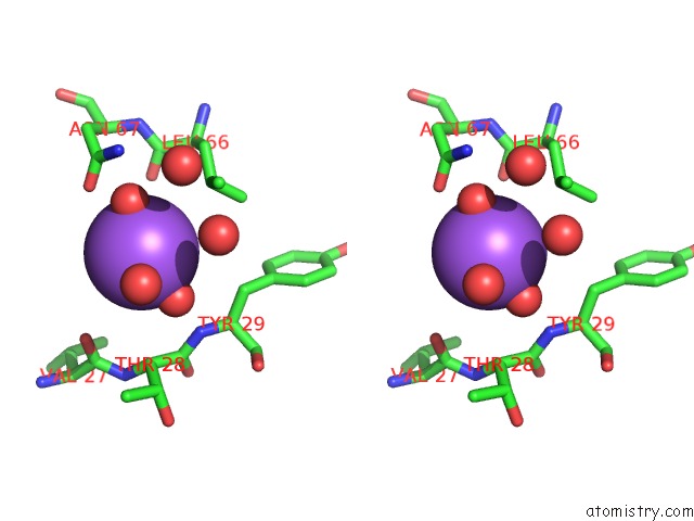
Stereo pair view
Mono view
Stereo pair view
A full contact list of Sodium with other atoms in the Na binding
site number 1 of Crystal Structure of the Urokinase within 5.0Å range:
|
Reference:
Y.N.Kang,
J.A.Stuckey,
V.Nienaber,
V.Giranda.
Crystal Structure of the Urokinase To Be Published.
Page generated: Mon Oct 7 15:28:23 2024
Last articles
Mg in 6H57Mg in 6H2N
Mg in 6H4C
Mg in 6H40
Mg in 6H2J
Mg in 6GZE
Mg in 6H21
Mg in 6H1J
Mg in 6H1V
Mg in 6H1M