Sodium »
PDB 4b1a-4bkr »
4bin »
Sodium in PDB 4bin: Crystal Structure of the E. Coli N-Acetylmuramoyl-L-Alanine Amidase Amic
Enzymatic activity of Crystal Structure of the E. Coli N-Acetylmuramoyl-L-Alanine Amidase Amic
All present enzymatic activity of Crystal Structure of the E. Coli N-Acetylmuramoyl-L-Alanine Amidase Amic:
3.5.1.28;
3.5.1.28;
Protein crystallography data
The structure of Crystal Structure of the E. Coli N-Acetylmuramoyl-L-Alanine Amidase Amic, PDB code: 4bin
was solved by
F.Kerff,
M.Rocaboy,
R.Herman,
E.Sauvage,
P.Charlier,
with X-Ray Crystallography technique. A brief refinement statistics is given in the table below:
| Resolution Low / High (Å) | 90.58 / 2.49 |
| Space group | P 21 21 21 |
| Cell size a, b, c (Å), α, β, γ (°) | 59.032, 68.442, 90.576, 90.00, 90.00, 90.00 |
| R / Rfree (%) | 17.564 / 23.101 |
Other elements in 4bin:
The structure of Crystal Structure of the E. Coli N-Acetylmuramoyl-L-Alanine Amidase Amic also contains other interesting chemical elements:
| Zinc | (Zn) | 1 atom |
Sodium Binding Sites:
The binding sites of Sodium atom in the Crystal Structure of the E. Coli N-Acetylmuramoyl-L-Alanine Amidase Amic
(pdb code 4bin). This binding sites where shown within
5.0 Angstroms radius around Sodium atom.
In total only one binding site of Sodium was determined in the Crystal Structure of the E. Coli N-Acetylmuramoyl-L-Alanine Amidase Amic, PDB code: 4bin:
In total only one binding site of Sodium was determined in the Crystal Structure of the E. Coli N-Acetylmuramoyl-L-Alanine Amidase Amic, PDB code: 4bin:
Sodium binding site 1 out of 1 in 4bin
Go back to
Sodium binding site 1 out
of 1 in the Crystal Structure of the E. Coli N-Acetylmuramoyl-L-Alanine Amidase Amic
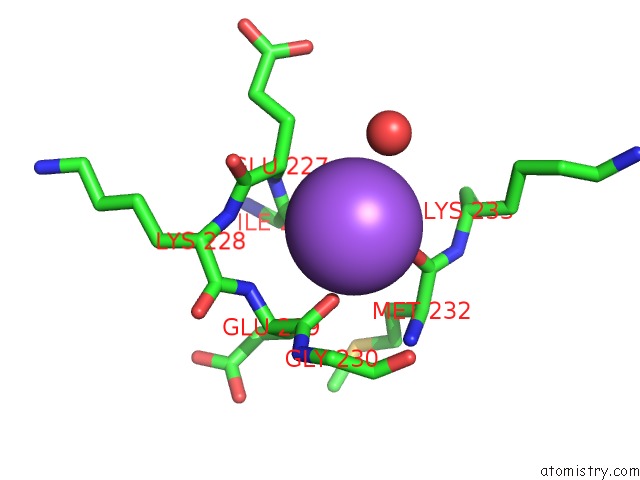
Mono view
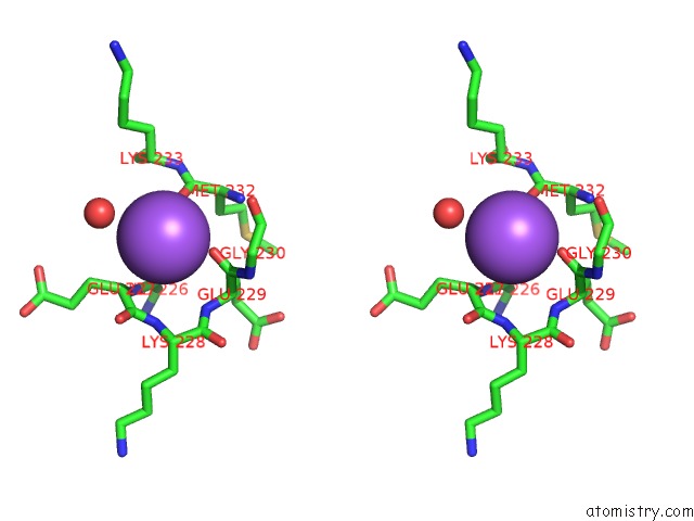
Stereo pair view
Mono view
Stereo pair view
A full contact list of Sodium with other atoms in the Na binding
site number 1 of Crystal Structure of the E. Coli N-Acetylmuramoyl-L-Alanine Amidase Amic within 5.0Å range:
|
Reference:
M.Rocaboy,
R.Herman,
E.Sauvage,
H.Remaut,
K.Moonens,
M.Terrak,
P.Charlier,
F.Kerff.
The Crystal Structure of the Cell Division Amidase Amic Reveals the Fold of the Amin Domain, A New Peptidoglycan Binding Domain. Mol.Microbiol. V. 90 267 2013.
ISSN: ISSN 0950-382X
PubMed: 23927005
DOI: 10.1111/MMI.12361
Page generated: Mon Oct 7 14:31:42 2024
ISSN: ISSN 0950-382X
PubMed: 23927005
DOI: 10.1111/MMI.12361
Last articles
Mn in 5NTGMn in 5NSQ
Mn in 5NRZ
Mn in 5NMP
Mn in 5NHA
Mn in 5NPK
Mn in 5NPP
Mn in 5NNB
Mn in 5NNA
Mn in 5NH9