Sodium »
PDB 3zux-4adj »
435d »
Sodium in PDB 435d: 5'-R(*Up*Ap*Gp*Cp*Cp*Cp*C)-3', 5'-R(*Gp*Gp*Gp*Gp*Cp*Up*A)-3'
Protein crystallography data
The structure of 5'-R(*Up*Ap*Gp*Cp*Cp*Cp*C)-3', 5'-R(*Gp*Gp*Gp*Gp*Cp*Up*A)-3', PDB code: 435d
was solved by
U.Mueller,
H.Schuebel,
M.Sprinzl,
U.Heinemann,
with X-Ray Crystallography technique. A brief refinement statistics is given in the table below:
| Resolution Low / High (Å) | 15.00 / 1.40 |
| Space group | P 1 |
| Cell size a, b, c (Å), α, β, γ (°) | 26.680, 26.697, 30.459, 104.29, 104.22, 91.66 |
| R / Rfree (%) | 15.8 / 21.7 |
Sodium Binding Sites:
The binding sites of Sodium atom in the 5'-R(*Up*Ap*Gp*Cp*Cp*Cp*C)-3', 5'-R(*Gp*Gp*Gp*Gp*Cp*Up*A)-3'
(pdb code 435d). This binding sites where shown within
5.0 Angstroms radius around Sodium atom.
In total 2 binding sites of Sodium where determined in the 5'-R(*Up*Ap*Gp*Cp*Cp*Cp*C)-3', 5'-R(*Gp*Gp*Gp*Gp*Cp*Up*A)-3', PDB code: 435d:
Jump to Sodium binding site number: 1; 2;
In total 2 binding sites of Sodium where determined in the 5'-R(*Up*Ap*Gp*Cp*Cp*Cp*C)-3', 5'-R(*Gp*Gp*Gp*Gp*Cp*Up*A)-3', PDB code: 435d:
Jump to Sodium binding site number: 1; 2;
Sodium binding site 1 out of 2 in 435d
Go back to
Sodium binding site 1 out
of 2 in the 5'-R(*Up*Ap*Gp*Cp*Cp*Cp*C)-3', 5'-R(*Gp*Gp*Gp*Gp*Cp*Up*A)-3'
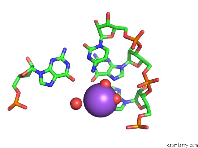
Mono view
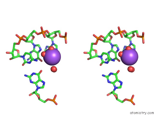
Stereo pair view
Mono view
Stereo pair view
A full contact list of Sodium with other atoms in the Na binding
site number 1 of 5'-R(*Up*Ap*Gp*Cp*Cp*Cp*C)-3', 5'-R(*Gp*Gp*Gp*Gp*Cp*Up*A)-3' within 5.0Å range:
|
Sodium binding site 2 out of 2 in 435d
Go back to
Sodium binding site 2 out
of 2 in the 5'-R(*Up*Ap*Gp*Cp*Cp*Cp*C)-3', 5'-R(*Gp*Gp*Gp*Gp*Cp*Up*A)-3'
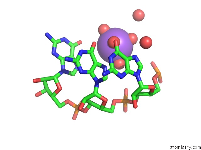
Mono view
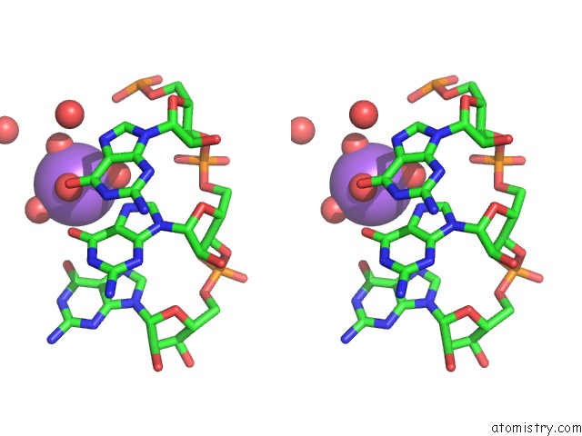
Stereo pair view
Mono view
Stereo pair view
A full contact list of Sodium with other atoms in the Na binding
site number 2 of 5'-R(*Up*Ap*Gp*Cp*Cp*Cp*C)-3', 5'-R(*Gp*Gp*Gp*Gp*Cp*Up*A)-3' within 5.0Å range:
|
Reference:
U.Mueller,
H.Schubel,
M.Sprinzl,
U.Heinemann.
Crystal Structure of Acceptor Stem of Trna(Ala) From Escherichia Coli Shows Unique G.U Wobble Base Pair at 1.16 A Resolution. Rna V. 5 670 1999.
ISSN: ISSN 1355-8382
PubMed: 10334337
DOI: 10.1017/S1355838299982304
Page generated: Mon Oct 7 14:16:09 2024
ISSN: ISSN 1355-8382
PubMed: 10334337
DOI: 10.1017/S1355838299982304
Last articles
K in 7NPXK in 7NWD
K in 7NXF
K in 7NF4
K in 7NHT
K in 7NKG
K in 7NF2
K in 7N9L
K in 7NFZ
K in 7NF3