Sodium »
PDB 3vif-3way »
3w8e »
Sodium in PDB 3w8e: Crystal Structure of D-3-Hydroxybutyrate Dehydrogenase From Alcaligenes Faecalis Complexed with Nad+ and A Substrate D-3- Hydroxybutyrate
Enzymatic activity of Crystal Structure of D-3-Hydroxybutyrate Dehydrogenase From Alcaligenes Faecalis Complexed with Nad+ and A Substrate D-3- Hydroxybutyrate
All present enzymatic activity of Crystal Structure of D-3-Hydroxybutyrate Dehydrogenase From Alcaligenes Faecalis Complexed with Nad+ and A Substrate D-3- Hydroxybutyrate:
1.1.1.30;
1.1.1.30;
Protein crystallography data
The structure of Crystal Structure of D-3-Hydroxybutyrate Dehydrogenase From Alcaligenes Faecalis Complexed with Nad+ and A Substrate D-3- Hydroxybutyrate, PDB code: 3w8e
was solved by
H.Kanazawa,
M.Tsunoda,
M.M.Hoque,
K.Suzuki,
T.Yamamoto,
A.Takenaka,
with X-Ray Crystallography technique. A brief refinement statistics is given in the table below:
| Resolution Low / High (Å) | 18.98 / 1.24 |
| Space group | P 42 21 2 |
| Cell size a, b, c (Å), α, β, γ (°) | 62.780, 62.780, 119.490, 90.00, 90.00, 90.00 |
| R / Rfree (%) | 16.5 / 19.2 |
Sodium Binding Sites:
The binding sites of Sodium atom in the Crystal Structure of D-3-Hydroxybutyrate Dehydrogenase From Alcaligenes Faecalis Complexed with Nad+ and A Substrate D-3- Hydroxybutyrate
(pdb code 3w8e). This binding sites where shown within
5.0 Angstroms radius around Sodium atom.
In total only one binding site of Sodium was determined in the Crystal Structure of D-3-Hydroxybutyrate Dehydrogenase From Alcaligenes Faecalis Complexed with Nad+ and A Substrate D-3- Hydroxybutyrate, PDB code: 3w8e:
In total only one binding site of Sodium was determined in the Crystal Structure of D-3-Hydroxybutyrate Dehydrogenase From Alcaligenes Faecalis Complexed with Nad+ and A Substrate D-3- Hydroxybutyrate, PDB code: 3w8e:
Sodium binding site 1 out of 1 in 3w8e
Go back to
Sodium binding site 1 out
of 1 in the Crystal Structure of D-3-Hydroxybutyrate Dehydrogenase From Alcaligenes Faecalis Complexed with Nad+ and A Substrate D-3- Hydroxybutyrate
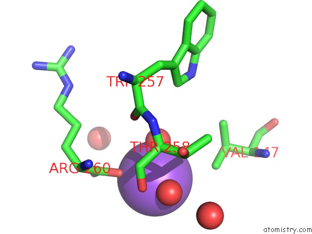
Mono view
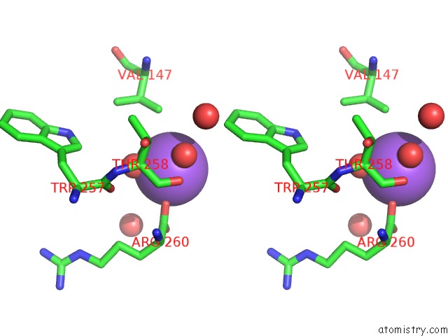
Stereo pair view
Mono view
Stereo pair view
A full contact list of Sodium with other atoms in the Na binding
site number 1 of Crystal Structure of D-3-Hydroxybutyrate Dehydrogenase From Alcaligenes Faecalis Complexed with Nad+ and A Substrate D-3- Hydroxybutyrate within 5.0Å range:
|
Reference:
H.Kanazawa,
M.Tsunoda,
M.M.Hoque,
K.Suzuki,
T.Yamamoto,
A.Takenaka.
High Resolution X-Ray Diffraction of D-3-Hydroxybutyrate Dehydrogenase From Alcaligenes Faecalis Complexed with Nad+ and D-3-Hydroxybutyrate To Be Published.
Page generated: Mon Oct 7 13:54:32 2024
Last articles
Fe in 6EYEFe in 6EXF
Fe in 6EX3
Fe in 6EX4
Fe in 6EUR
Fe in 6EX0
Fe in 6EUO
Fe in 6EWZ
Fe in 6ELQ
Fe in 6ETB