Sodium »
PDB 3v45-3vi2 »
3vdg »
Sodium in PDB 3vdg: Crystal Structure of Enolase MSMEG_6132 (Target Efi-502282) From Mycobacterium Smegmatis Str. MC2 155 Complexed with Formate and Acetate
Protein crystallography data
The structure of Crystal Structure of Enolase MSMEG_6132 (Target Efi-502282) From Mycobacterium Smegmatis Str. MC2 155 Complexed with Formate and Acetate, PDB code: 3vdg
was solved by
Y.Patskovsky,
R.Toro,
R.Bhosle,
B.Hillerich,
R.D.Seidel,
E.Washington,
A.Scott Glenn,
S.Chowdhury,
B.Evans,
J.Hammonds,
W.D.Zencheck,
H.J.Imker,
J.A.Gerlt,
S.C.Almo,
Enzyme Function Initiative (Efi),
with X-Ray Crystallography technique. A brief refinement statistics is given in the table below:
| Resolution Low / High (Å) | 50.00 / 1.90 |
| Space group | P 61 2 2 |
| Cell size a, b, c (Å), α, β, γ (°) | 94.949, 94.949, 259.752, 90.00, 90.00, 120.00 |
| R / Rfree (%) | 15.9 / 18.6 |
Other elements in 3vdg:
The structure of Crystal Structure of Enolase MSMEG_6132 (Target Efi-502282) From Mycobacterium Smegmatis Str. MC2 155 Complexed with Formate and Acetate also contains other interesting chemical elements:
| Chlorine | (Cl) | 1 atom |
Sodium Binding Sites:
The binding sites of Sodium atom in the Crystal Structure of Enolase MSMEG_6132 (Target Efi-502282) From Mycobacterium Smegmatis Str. MC2 155 Complexed with Formate and Acetate
(pdb code 3vdg). This binding sites where shown within
5.0 Angstroms radius around Sodium atom.
In total only one binding site of Sodium was determined in the Crystal Structure of Enolase MSMEG_6132 (Target Efi-502282) From Mycobacterium Smegmatis Str. MC2 155 Complexed with Formate and Acetate, PDB code: 3vdg:
In total only one binding site of Sodium was determined in the Crystal Structure of Enolase MSMEG_6132 (Target Efi-502282) From Mycobacterium Smegmatis Str. MC2 155 Complexed with Formate and Acetate, PDB code: 3vdg:
Sodium binding site 1 out of 1 in 3vdg
Go back to
Sodium binding site 1 out
of 1 in the Crystal Structure of Enolase MSMEG_6132 (Target Efi-502282) From Mycobacterium Smegmatis Str. MC2 155 Complexed with Formate and Acetate
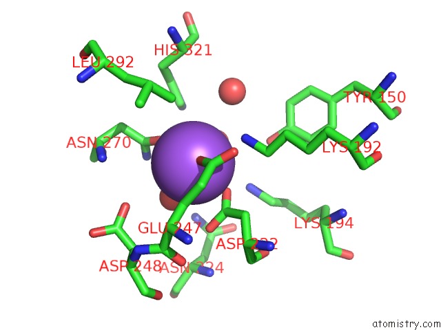
Mono view
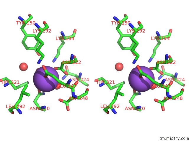
Stereo pair view
Mono view
Stereo pair view
A full contact list of Sodium with other atoms in the Na binding
site number 1 of Crystal Structure of Enolase MSMEG_6132 (Target Efi-502282) From Mycobacterium Smegmatis Str. MC2 155 Complexed with Formate and Acetate within 5.0Å range:
|
Reference:
Y.Patskovsky,
R.Toro,
R.Bhosle,
B.Hillerich,
R.D.Seidel,
E.Washington,
A.Scott Glenn,
S.Chowdhury,
B.Evans,
J.Hammonds,
W.D.Zencheck,
H.J.Imker,
J.A.Gerlt,
S.C.Almo.
Crystal Structure of Enolase MSMEG_6132 From Mycobacterium Smegmatis To Be Published.
Page generated: Mon Oct 7 13:47:52 2024
Last articles
Mg in 2AG1Mg in 2AG0
Mg in 2AET
Mg in 2AER
Mg in 2AEL
Mg in 2AEK
Mg in 2AE6
Mg in 2AD5
Mg in 2ACX
Mg in 2A9R