Sodium »
PDB 3v45-3vi2 »
3va7 »
Sodium in PDB 3va7: Crystal Structure of the Kluyveromyces Lactis Urea Carboxylase
Enzymatic activity of Crystal Structure of the Kluyveromyces Lactis Urea Carboxylase
All present enzymatic activity of Crystal Structure of the Kluyveromyces Lactis Urea Carboxylase:
6.3.4.6;
6.3.4.6;
Protein crystallography data
The structure of Crystal Structure of the Kluyveromyces Lactis Urea Carboxylase, PDB code: 3va7
was solved by
C.Fan,
S.Xiang,
with X-Ray Crystallography technique. A brief refinement statistics is given in the table below:
| Resolution Low / High (Å) | 50.00 / 2.60 |
| Space group | P 43 21 2 |
| Cell size a, b, c (Å), α, β, γ (°) | 126.660, 126.660, 217.870, 90.00, 90.00, 90.00 |
| R / Rfree (%) | 18.7 / 25.5 |
Sodium Binding Sites:
The binding sites of Sodium atom in the Crystal Structure of the Kluyveromyces Lactis Urea Carboxylase
(pdb code 3va7). This binding sites where shown within
5.0 Angstroms radius around Sodium atom.
In total only one binding site of Sodium was determined in the Crystal Structure of the Kluyveromyces Lactis Urea Carboxylase, PDB code: 3va7:
In total only one binding site of Sodium was determined in the Crystal Structure of the Kluyveromyces Lactis Urea Carboxylase, PDB code: 3va7:
Sodium binding site 1 out of 1 in 3va7
Go back to
Sodium binding site 1 out
of 1 in the Crystal Structure of the Kluyveromyces Lactis Urea Carboxylase
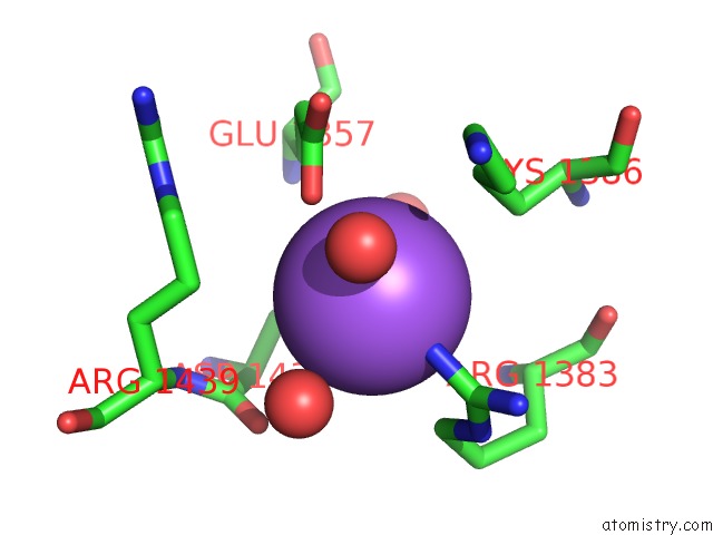
Mono view
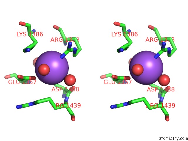
Stereo pair view
Mono view
Stereo pair view
A full contact list of Sodium with other atoms in the Na binding
site number 1 of Crystal Structure of the Kluyveromyces Lactis Urea Carboxylase within 5.0Å range:
|
Reference:
C.Fan,
C.Y.Chou,
L.Tong,
S.Xiang.
Crystal Structure of Urea Carboxylase Provides Insights Into the Carboxyltransfer Reaction J.Biol.Chem. V. 287 9389 2012.
ISSN: ISSN 0021-9258
PubMed: 22277658
DOI: 10.1074/JBC.M111.319475
Page generated: Mon Oct 7 13:39:51 2024
ISSN: ISSN 0021-9258
PubMed: 22277658
DOI: 10.1074/JBC.M111.319475
Last articles
Mg in 2AETMg in 2AER
Mg in 2AEL
Mg in 2AEK
Mg in 2AE6
Mg in 2AD5
Mg in 2ACX
Mg in 2A9R
Mg in 2A9K
Mg in 2A9F