Sodium »
PDB 3uq0-3v3r »
3usj »
Sodium in PDB 3usj: Crystal Structure of Leut Bound to L-Leucine in Space Group P21 From Lipid Bicelles
Protein crystallography data
The structure of Crystal Structure of Leut Bound to L-Leucine in Space Group P21 From Lipid Bicelles, PDB code: 3usj
was solved by
H.Wang,
J.Elferich,
E.Gouaux,
with X-Ray Crystallography technique. A brief refinement statistics is given in the table below:
| Resolution Low / High (Å) | 48.33 / 3.50 |
| Space group | P 1 21 1 |
| Cell size a, b, c (Å), α, β, γ (°) | 57.307, 179.880, 57.207, 90.00, 89.96, 90.00 |
| R / Rfree (%) | 26.4 / 30.9 |
Sodium Binding Sites:
The binding sites of Sodium atom in the Crystal Structure of Leut Bound to L-Leucine in Space Group P21 From Lipid Bicelles
(pdb code 3usj). This binding sites where shown within
5.0 Angstroms radius around Sodium atom.
In total 4 binding sites of Sodium where determined in the Crystal Structure of Leut Bound to L-Leucine in Space Group P21 From Lipid Bicelles, PDB code: 3usj:
Jump to Sodium binding site number: 1; 2; 3; 4;
In total 4 binding sites of Sodium where determined in the Crystal Structure of Leut Bound to L-Leucine in Space Group P21 From Lipid Bicelles, PDB code: 3usj:
Jump to Sodium binding site number: 1; 2; 3; 4;
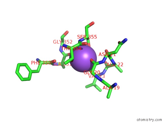
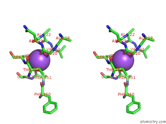
Sodium binding site 1 out of 4 in 3usj
Go back to
Sodium binding site 1 out
of 4 in the Crystal Structure of Leut Bound to L-Leucine in Space Group P21 From Lipid Bicelles
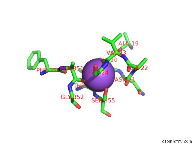
Mono view
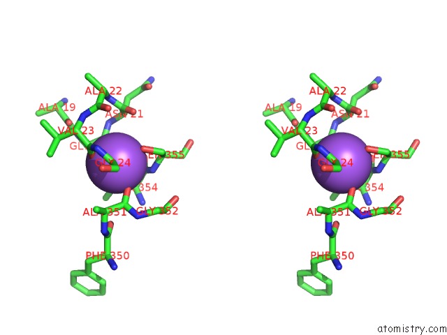
Stereo pair view
Mono view
Stereo pair view
A full contact list of Sodium with other atoms in the Na binding
site number 1 of Crystal Structure of Leut Bound to L-Leucine in Space Group P21 From Lipid Bicelles within 5.0Å range:
|
Sodium binding site 2 out of 4 in 3usj
Go back to
Sodium binding site 2 out
of 4 in the Crystal Structure of Leut Bound to L-Leucine in Space Group P21 From Lipid Bicelles
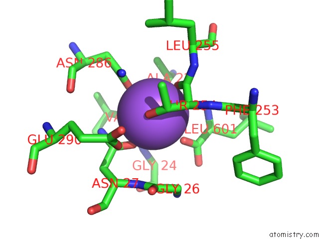
Mono view
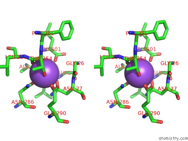
Stereo pair view
Mono view
Stereo pair view
A full contact list of Sodium with other atoms in the Na binding
site number 2 of Crystal Structure of Leut Bound to L-Leucine in Space Group P21 From Lipid Bicelles within 5.0Å range:
|
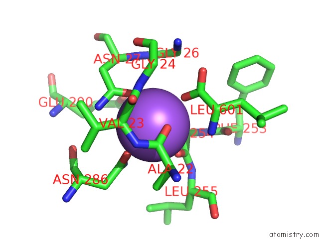
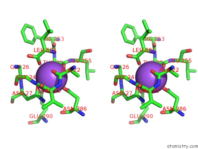
Sodium binding site 3 out of 4 in 3usj
Go back to
Sodium binding site 3 out
of 4 in the Crystal Structure of Leut Bound to L-Leucine in Space Group P21 From Lipid Bicelles
Mono view
Stereo pair view
Mono view
Stereo pair view
A full contact list of Sodium with other atoms in the Na binding
site number 3 of Crystal Structure of Leut Bound to L-Leucine in Space Group P21 From Lipid Bicelles within 5.0Å range:
|
Sodium binding site 4 out of 4 in 3usj
Go back to
Sodium binding site 4 out
of 4 in the Crystal Structure of Leut Bound to L-Leucine in Space Group P21 From Lipid Bicelles
Mono view
Stereo pair view
Mono view
Stereo pair view
A full contact list of Sodium with other atoms in the Na binding
site number 4 of Crystal Structure of Leut Bound to L-Leucine in Space Group P21 From Lipid Bicelles within 5.0Å range:
|
Reference:
H.Wang,
J.Elferich,
E.Gouaux.
Structures of Leut in Bicelles Define Conformation and Substrate Binding in A Membrane-Like Context. Nat.Struct.Mol.Biol. V. 19 212 2012.
ISSN: ISSN 1545-9993
PubMed: 22245965
DOI: 10.1038/NSMB.2215
Page generated: Mon Oct 7 13:32:02 2024
ISSN: ISSN 1545-9993
PubMed: 22245965
DOI: 10.1038/NSMB.2215
Last articles
K in 6KSHK in 6L5G
K in 6L32
K in 6KY3
K in 6KFL
K in 6KKT
K in 6JXK
K in 6JXH
K in 6KKR
K in 6K1K