Sodium »
PDB 3u0a-3upw »
3ud2 »
Sodium in PDB 3ud2: Crystal Structure of Selenomethionine ZU5A-ZU5B Protein Domains of Human Erythrocyte Ankyrin
Protein crystallography data
The structure of Crystal Structure of Selenomethionine ZU5A-ZU5B Protein Domains of Human Erythrocyte Ankyrin, PDB code: 3ud2
was solved by
M.Yasunaga,
J.J.Ipsaro,
A.Mondragon,
with X-Ray Crystallography technique. A brief refinement statistics is given in the table below:
| Resolution Low / High (Å) | 24.90 / 2.21 |
| Space group | C 1 2 1 |
| Cell size a, b, c (Å), α, β, γ (°) | 279.480, 40.873, 95.249, 90.00, 92.01, 90.00 |
| R / Rfree (%) | 22.1 / 25.9 |
Other elements in 3ud2:
The structure of Crystal Structure of Selenomethionine ZU5A-ZU5B Protein Domains of Human Erythrocyte Ankyrin also contains other interesting chemical elements:
| Chlorine | (Cl) | 1 atom |
Sodium Binding Sites:
Pages:
>>> Page 1 <<< Page 2, Binding sites: 11 - 17;Binding sites:
The binding sites of Sodium atom in the Crystal Structure of Selenomethionine ZU5A-ZU5B Protein Domains of Human Erythrocyte Ankyrin (pdb code 3ud2). This binding sites where shown within 5.0 Angstroms radius around Sodium atom.In total 17 binding sites of Sodium where determined in the Crystal Structure of Selenomethionine ZU5A-ZU5B Protein Domains of Human Erythrocyte Ankyrin, PDB code: 3ud2:
Jump to Sodium binding site number: 1; 2; 3; 4; 5; 6; 7; 8; 9; 10;
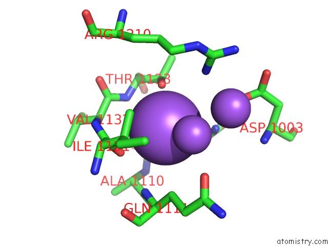
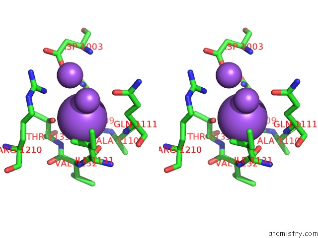
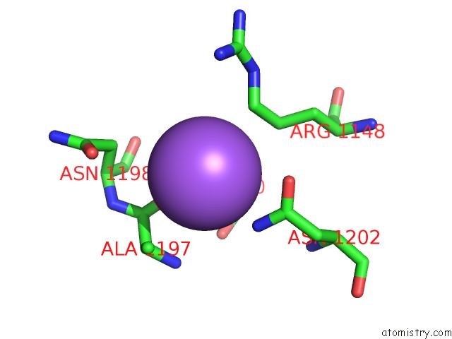
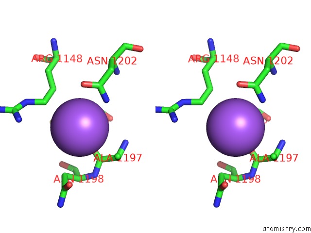
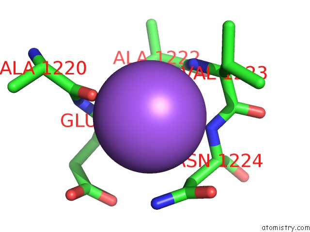
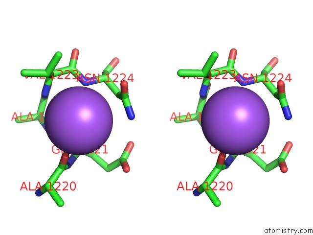
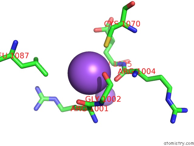
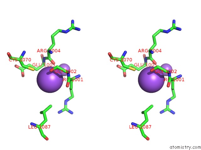
Sodium binding site 1 out of 17 in 3ud2
Go back to
Sodium binding site 1 out
of 17 in the Crystal Structure of Selenomethionine ZU5A-ZU5B Protein Domains of Human Erythrocyte Ankyrin
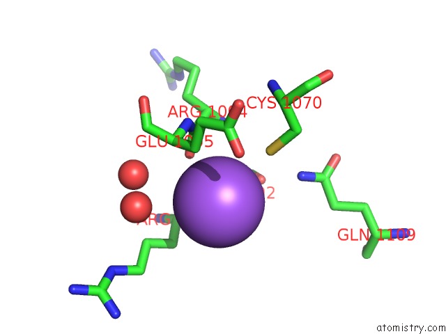
Mono view
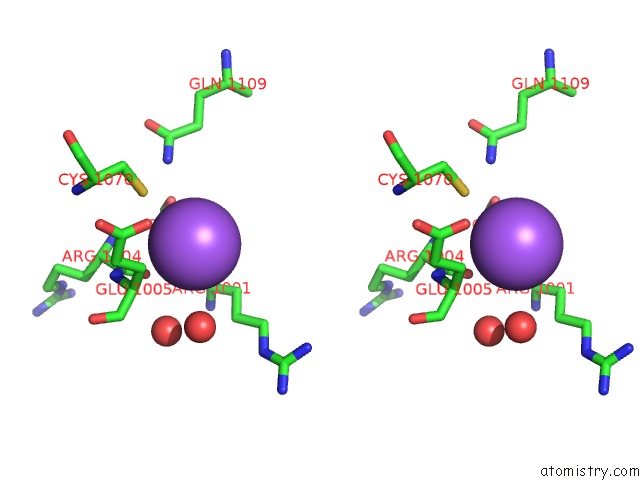
Stereo pair view
Mono view
Stereo pair view
A full contact list of Sodium with other atoms in the Na binding
site number 1 of Crystal Structure of Selenomethionine ZU5A-ZU5B Protein Domains of Human Erythrocyte Ankyrin within 5.0Å range:
|
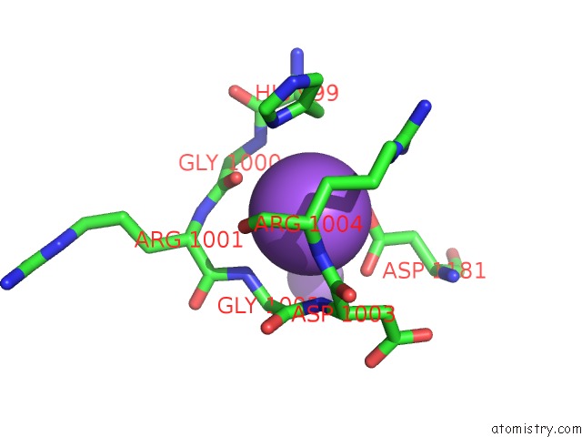
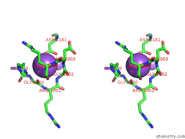
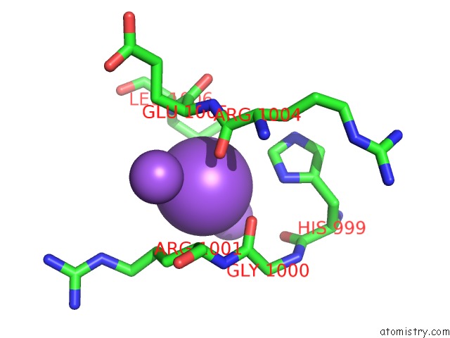
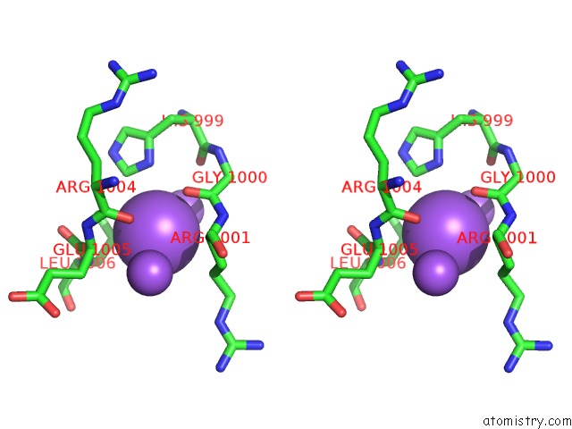
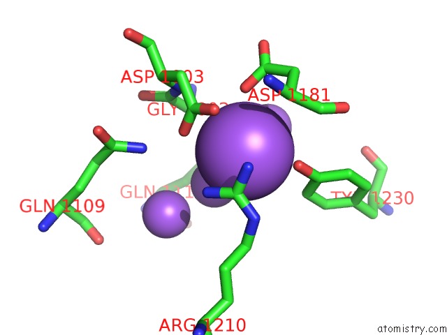
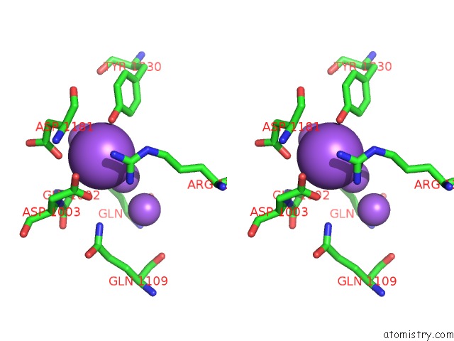
Sodium binding site 2 out of 17 in 3ud2
Go back to
Sodium binding site 2 out
of 17 in the Crystal Structure of Selenomethionine ZU5A-ZU5B Protein Domains of Human Erythrocyte Ankyrin
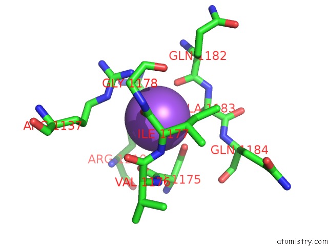
Mono view
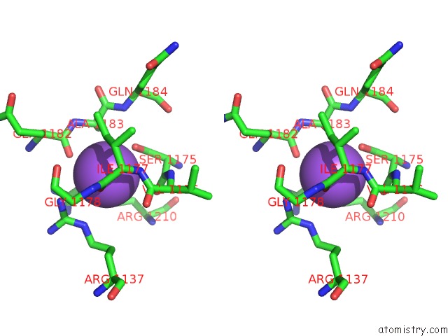
Stereo pair view
Mono view
Stereo pair view
A full contact list of Sodium with other atoms in the Na binding
site number 2 of Crystal Structure of Selenomethionine ZU5A-ZU5B Protein Domains of Human Erythrocyte Ankyrin within 5.0Å range:
|
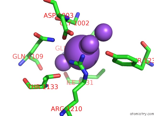
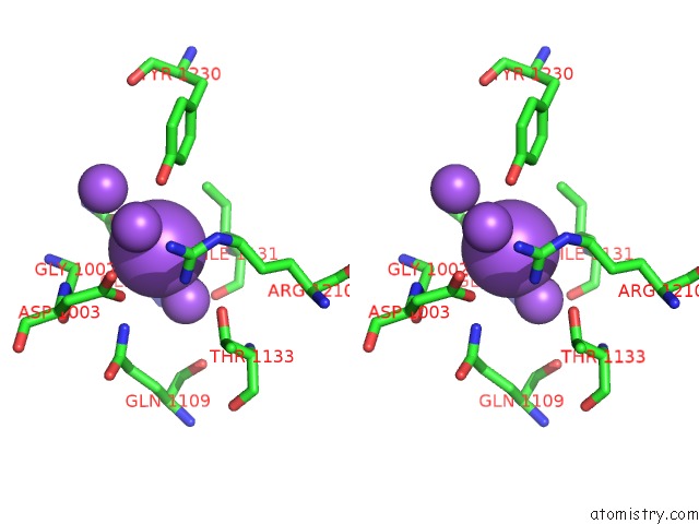
Sodium binding site 3 out of 17 in 3ud2
Go back to
Sodium binding site 3 out
of 17 in the Crystal Structure of Selenomethionine ZU5A-ZU5B Protein Domains of Human Erythrocyte Ankyrin
Mono view
Stereo pair view
Mono view
Stereo pair view
A full contact list of Sodium with other atoms in the Na binding
site number 3 of Crystal Structure of Selenomethionine ZU5A-ZU5B Protein Domains of Human Erythrocyte Ankyrin within 5.0Å range:
|
Sodium binding site 4 out of 17 in 3ud2
Go back to
Sodium binding site 4 out
of 17 in the Crystal Structure of Selenomethionine ZU5A-ZU5B Protein Domains of Human Erythrocyte Ankyrin
Mono view
Stereo pair view
Mono view
Stereo pair view
A full contact list of Sodium with other atoms in the Na binding
site number 4 of Crystal Structure of Selenomethionine ZU5A-ZU5B Protein Domains of Human Erythrocyte Ankyrin within 5.0Å range:
|
Sodium binding site 5 out of 17 in 3ud2
Go back to
Sodium binding site 5 out
of 17 in the Crystal Structure of Selenomethionine ZU5A-ZU5B Protein Domains of Human Erythrocyte Ankyrin
Mono view
Stereo pair view
Mono view
Stereo pair view
A full contact list of Sodium with other atoms in the Na binding
site number 5 of Crystal Structure of Selenomethionine ZU5A-ZU5B Protein Domains of Human Erythrocyte Ankyrin within 5.0Å range:
|
Sodium binding site 6 out of 17 in 3ud2
Go back to
Sodium binding site 6 out
of 17 in the Crystal Structure of Selenomethionine ZU5A-ZU5B Protein Domains of Human Erythrocyte Ankyrin
Mono view
Stereo pair view
Mono view
Stereo pair view
A full contact list of Sodium with other atoms in the Na binding
site number 6 of Crystal Structure of Selenomethionine ZU5A-ZU5B Protein Domains of Human Erythrocyte Ankyrin within 5.0Å range:
|
Sodium binding site 7 out of 17 in 3ud2
Go back to
Sodium binding site 7 out
of 17 in the Crystal Structure of Selenomethionine ZU5A-ZU5B Protein Domains of Human Erythrocyte Ankyrin
Mono view
Stereo pair view
Mono view
Stereo pair view
A full contact list of Sodium with other atoms in the Na binding
site number 7 of Crystal Structure of Selenomethionine ZU5A-ZU5B Protein Domains of Human Erythrocyte Ankyrin within 5.0Å range:
|
Sodium binding site 8 out of 17 in 3ud2
Go back to
Sodium binding site 8 out
of 17 in the Crystal Structure of Selenomethionine ZU5A-ZU5B Protein Domains of Human Erythrocyte Ankyrin
Mono view
Stereo pair view
Mono view
Stereo pair view
A full contact list of Sodium with other atoms in the Na binding
site number 8 of Crystal Structure of Selenomethionine ZU5A-ZU5B Protein Domains of Human Erythrocyte Ankyrin within 5.0Å range:
|
Sodium binding site 9 out of 17 in 3ud2
Go back to
Sodium binding site 9 out
of 17 in the Crystal Structure of Selenomethionine ZU5A-ZU5B Protein Domains of Human Erythrocyte Ankyrin
Mono view
Stereo pair view
Mono view
Stereo pair view
A full contact list of Sodium with other atoms in the Na binding
site number 9 of Crystal Structure of Selenomethionine ZU5A-ZU5B Protein Domains of Human Erythrocyte Ankyrin within 5.0Å range:
|
Sodium binding site 10 out of 17 in 3ud2
Go back to
Sodium binding site 10 out
of 17 in the Crystal Structure of Selenomethionine ZU5A-ZU5B Protein Domains of Human Erythrocyte Ankyrin
Mono view
Stereo pair view
Mono view
Stereo pair view
A full contact list of Sodium with other atoms in the Na binding
site number 10 of Crystal Structure of Selenomethionine ZU5A-ZU5B Protein Domains of Human Erythrocyte Ankyrin within 5.0Å range:
|
Reference:
M.Yasunaga,
J.J.Ipsaro,
A.Mondragon.
Structurally Similar But Functionally Diverse ZU5 Domains in Human Erythrocyte Ankyrin. J.Mol.Biol. V. 417 336 2012.
ISSN: ISSN 0022-2836
PubMed: 22310050
DOI: 10.1016/J.JMB.2012.01.041
Page generated: Mon Oct 7 13:27:06 2024
ISSN: ISSN 0022-2836
PubMed: 22310050
DOI: 10.1016/J.JMB.2012.01.041
Last articles
Mg in 1L9BMg in 1L8T
Mg in 1L8A
Mg in 1L8Q
Mg in 1L7N
Mg in 1L7M
Mg in 1L6Y
Mg in 1L6S
Mg in 1L5Y
Mg in 1L5U