Sodium »
PDB 3sib-3t09 »
3sib »
Sodium in PDB 3sib: Crystal Structure of URE3-Binding Protein, Wild-Type
Protein crystallography data
The structure of Crystal Structure of URE3-Binding Protein, Wild-Type, PDB code: 3sib
was solved by
Seattle Structural Genomics Center For Infectious Disease (Ssgcid),
with X-Ray Crystallography technique. A brief refinement statistics is given in the table below:
| Resolution Low / High (Å) | 41.16 / 1.90 |
| Space group | I 4 2 2 |
| Cell size a, b, c (Å), α, β, γ (°) | 99.699, 99.699, 107.034, 90.00, 90.00, 90.00 |
| R / Rfree (%) | 15.9 / 19 |
Other elements in 3sib:
The structure of Crystal Structure of URE3-Binding Protein, Wild-Type also contains other interesting chemical elements:
| Calcium | (Ca) | 2 atoms |
Sodium Binding Sites:
The binding sites of Sodium atom in the Crystal Structure of URE3-Binding Protein, Wild-Type
(pdb code 3sib). This binding sites where shown within
5.0 Angstroms radius around Sodium atom.
In total only one binding site of Sodium was determined in the Crystal Structure of URE3-Binding Protein, Wild-Type, PDB code: 3sib:
In total only one binding site of Sodium was determined in the Crystal Structure of URE3-Binding Protein, Wild-Type, PDB code: 3sib:
Sodium binding site 1 out of 1 in 3sib
Go back to
Sodium binding site 1 out
of 1 in the Crystal Structure of URE3-Binding Protein, Wild-Type
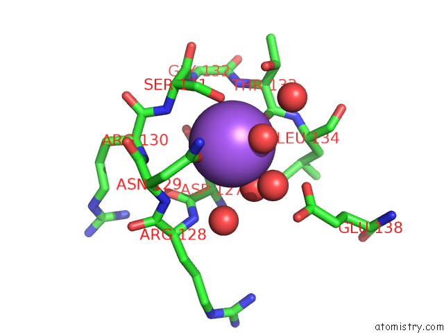
Mono view
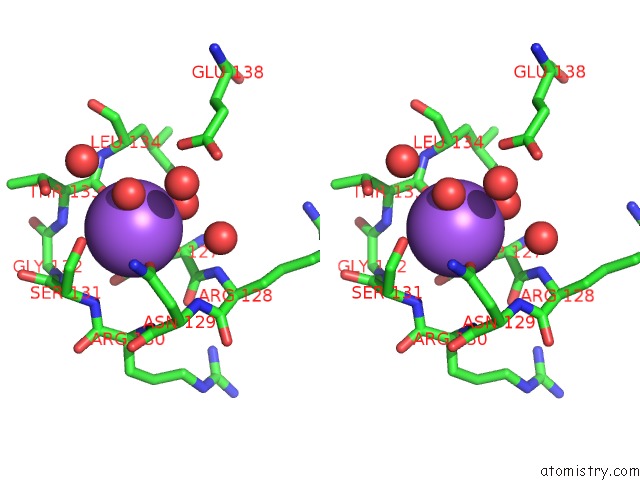
Stereo pair view
Mono view
Stereo pair view
A full contact list of Sodium with other atoms in the Na binding
site number 1 of Crystal Structure of URE3-Binding Protein, Wild-Type within 5.0Å range:
|
Reference:
Seattle Structural Genomics Center For Infectious Disease(Ssgcid),
A.Gardberg,
T.Edwards,
B.Staker,
P.Skubak,
C.Gilchrist,
L.Stewart.
Crystal Structure of URE3-Binding Protein To Be Published.
Page generated: Mon Oct 7 12:58:23 2024
Last articles
K in 5AVSK in 5AOH
K in 5AOP
K in 5AMM
K in 5A8R
K in 5ALA
K in 5AH1
K in 5AL9
K in 5AF2
K in 5AH0