Sodium »
PDB 3rnx-3si4 »
3rrx »
Sodium in PDB 3rrx: Crystal Structure of Q683A Mutant of Exo-1,3/1,4-Beta-Glucanase (Exop) From Pseudoalteromonas Sp. BB1
Protein crystallography data
The structure of Crystal Structure of Q683A Mutant of Exo-1,3/1,4-Beta-Glucanase (Exop) From Pseudoalteromonas Sp. BB1, PDB code: 3rrx
was solved by
Y.Nakatani,
S.M.Cutfield,
J.F.Cutfield,
with X-Ray Crystallography technique. A brief refinement statistics is given in the table below:
| Resolution Low / High (Å) | 19.98 / 1.90 |
| Space group | C 2 2 21 |
| Cell size a, b, c (Å), α, β, γ (°) | 68.247, 178.601, 176.047, 90.00, 90.00, 90.00 |
| R / Rfree (%) | 16.2 / 19 |
Other elements in 3rrx:
The structure of Crystal Structure of Q683A Mutant of Exo-1,3/1,4-Beta-Glucanase (Exop) From Pseudoalteromonas Sp. BB1 also contains other interesting chemical elements:
| Calcium | (Ca) | 1 atom |
Sodium Binding Sites:
The binding sites of Sodium atom in the Crystal Structure of Q683A Mutant of Exo-1,3/1,4-Beta-Glucanase (Exop) From Pseudoalteromonas Sp. BB1
(pdb code 3rrx). This binding sites where shown within
5.0 Angstroms radius around Sodium atom.
In total only one binding site of Sodium was determined in the Crystal Structure of Q683A Mutant of Exo-1,3/1,4-Beta-Glucanase (Exop) From Pseudoalteromonas Sp. BB1, PDB code: 3rrx:
In total only one binding site of Sodium was determined in the Crystal Structure of Q683A Mutant of Exo-1,3/1,4-Beta-Glucanase (Exop) From Pseudoalteromonas Sp. BB1, PDB code: 3rrx:
Sodium binding site 1 out of 1 in 3rrx
Go back to
Sodium binding site 1 out
of 1 in the Crystal Structure of Q683A Mutant of Exo-1,3/1,4-Beta-Glucanase (Exop) From Pseudoalteromonas Sp. BB1
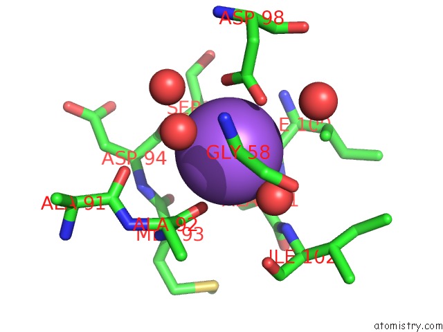
Mono view
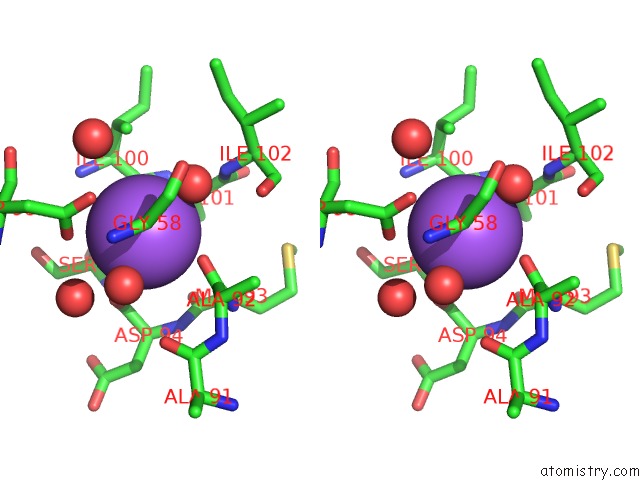
Stereo pair view
Mono view
Stereo pair view
A full contact list of Sodium with other atoms in the Na binding
site number 1 of Crystal Structure of Q683A Mutant of Exo-1,3/1,4-Beta-Glucanase (Exop) From Pseudoalteromonas Sp. BB1 within 5.0Å range:
|
Reference:
Y.Nakatani,
S.M.Cutfield,
N.P.Cowieson,
J.F.Cutfield.
Structure and Activity of Exo-1,3/1,4-Beta-Glucanase From Marine Bacterium Pseudoalteromonas Sp. BB1 Showing A Novel C-Terminal Domain Febs J. V. 279 464 2012.
ISSN: ISSN 1742-464X
PubMed: 22129429
DOI: 10.1111/J.1742-4658.2011.08439.X
Page generated: Mon Oct 7 12:51:54 2024
ISSN: ISSN 1742-464X
PubMed: 22129429
DOI: 10.1111/J.1742-4658.2011.08439.X
Last articles
Mg in 4LCZMg in 4LF2
Mg in 4LF1
Mg in 4LEM
Mg in 4LCK
Mg in 4LE0
Mg in 4LDZ
Mg in 4LDT
Mg in 4LA7
Mg in 4LDJ